Copyright
©The Author(s) 2015.
World J Gastroenterol. Mar 14, 2015; 21(10): 3139-3145
Published online Mar 14, 2015. doi: 10.3748/wjg.v21.i10.3139
Published online Mar 14, 2015. doi: 10.3748/wjg.v21.i10.3139
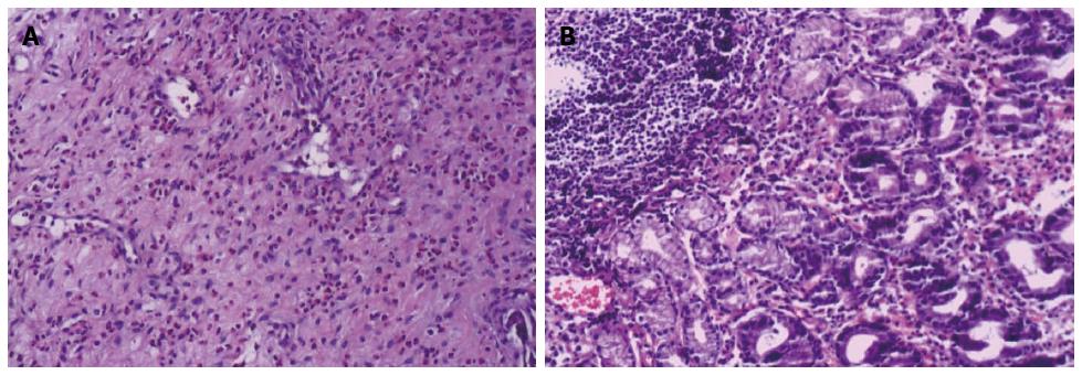
Figure 3 Histopathological examination of case 1.
A: Histopathological examination of the specimen obtained by cystoscopy showed infiltration of the bladder wall mucosa and submucosa by numerous eosinophils (HE staining, × 200); B: Extensive subserosal eosinophilic infiltration was also demonstrated by gastric and duodenal biopsies (HE staining, × 400).
- Citation: Han SG, Chen Y, Qian ZH, Yang L, Yu RS, Zhu XL, Li QH, Chen Q. Eosinophilic gastroenteritis associated with eosinophilic cystitis: Computed tomography and magnetic resonance imaging findings. World J Gastroenterol 2015; 21(10): 3139-3145
- URL: https://www.wjgnet.com/1007-9327/full/v21/i10/3139.htm
- DOI: https://dx.doi.org/10.3748/wjg.v21.i10.3139


