Copyright
©The Author(s) 2015.
World J Gastroenterol. Mar 14, 2015; 21(10): 3139-3145
Published online Mar 14, 2015. doi: 10.3748/wjg.v21.i10.3139
Published online Mar 14, 2015. doi: 10.3748/wjg.v21.i10.3139
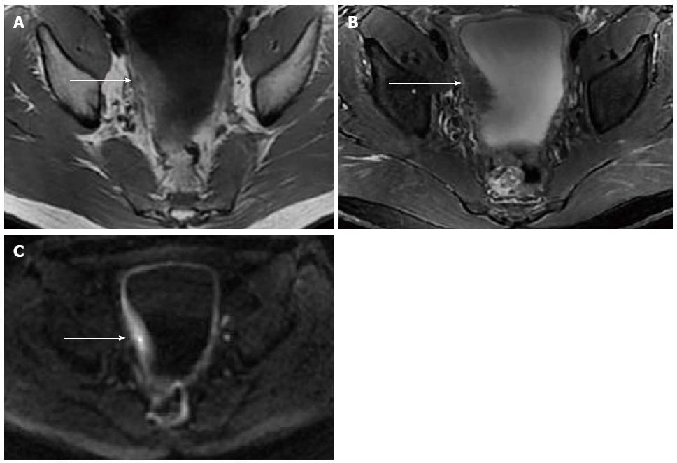
Figure 2 Pelvic magnetic resonance imaging of case 1.
Pelvic magnetic resonance imaging revealed thickening of the bladder wall measuring 18 mm in thickness of the right part of the bladder wall (arrow). The thickened bladder wall exhibited mildly homogeneous hyper-intensity relative to muscle on T1WI (A), hypo-intensity on fat-suppressed T2WI (B), and slight hyper-intensity on DWI (b-value = 1000) (C).
- Citation: Han SG, Chen Y, Qian ZH, Yang L, Yu RS, Zhu XL, Li QH, Chen Q. Eosinophilic gastroenteritis associated with eosinophilic cystitis: Computed tomography and magnetic resonance imaging findings. World J Gastroenterol 2015; 21(10): 3139-3145
- URL: https://www.wjgnet.com/1007-9327/full/v21/i10/3139.htm
- DOI: https://dx.doi.org/10.3748/wjg.v21.i10.3139


