Copyright
©The Author(s) 2015.
World J Gastroenterol. Mar 14, 2015; 21(10): 3139-3145
Published online Mar 14, 2015. doi: 10.3748/wjg.v21.i10.3139
Published online Mar 14, 2015. doi: 10.3748/wjg.v21.i10.3139
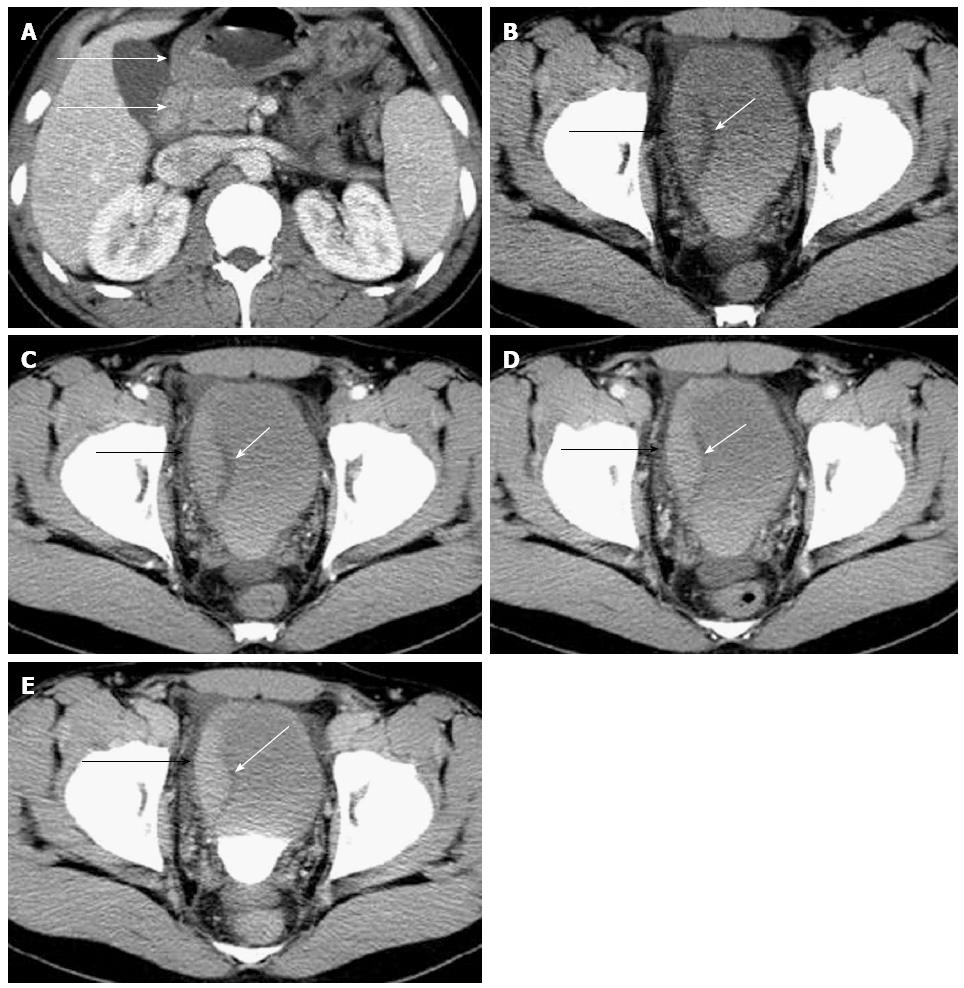
Figure 1 Contrast-enhanced computed tomography of case 1.
Contrast-enhanced computed tomography (CT) showed the wall thickening of gastric antrum (white arrow) and duodenum (white arrow) with inhomogeneous reinforcement (A). Contrast-enhanced CT examination also showed coexistent right lateral bladder wall asymmetrical thickening with progressive enhancement (black arrow) on the plain (B), arterial (C), portal (D), and delayed phase (E) scans, with preservation of the low-density mucosal line (white arrow).
- Citation: Han SG, Chen Y, Qian ZH, Yang L, Yu RS, Zhu XL, Li QH, Chen Q. Eosinophilic gastroenteritis associated with eosinophilic cystitis: Computed tomography and magnetic resonance imaging findings. World J Gastroenterol 2015; 21(10): 3139-3145
- URL: https://www.wjgnet.com/1007-9327/full/v21/i10/3139.htm
- DOI: https://dx.doi.org/10.3748/wjg.v21.i10.3139


