Copyright
©2014 Baishideng Publishing Group Inc.
World J Gastroenterol. Dec 21, 2014; 20(47): 17804-17818
Published online Dec 21, 2014. doi: 10.3748/wjg.v20.i47.17804
Published online Dec 21, 2014. doi: 10.3748/wjg.v20.i47.17804
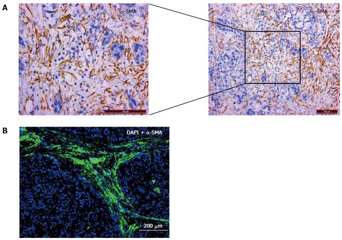
Figure 1 Hepatocellular carcinoma-associated cancer-associated fibroblasts and hepatocellular carcinoma cells.
A: The distribution of H-CAFs identified by α-SMA (+) expression in a HCC specimen is detected by immunohistochemistry. Expression of α-SMA (shown in brown color) is detected to confirm the presence of H-CAFs, which are abundant in tumor tissue; B: The presence of H-CAFs in HCC tissue demonstrated by immunofluorescence. The blue color indicates HCC nucleus, and the green H-CAFs with α-SMA stained. CAFs are circulating the cancer nests in the malignant tissue. HCC: Hepatocellular carcinoma; H-CAF: HCC-associated cancer-associated fibroblasts; α-SMA: α-smooth muscle actin.
- Citation: Huang L, Xu AM, Liu S, Liu W, Li TJ. Cancer-associated fibroblasts in digestive tumors. World J Gastroenterol 2014; 20(47): 17804-17818
- URL: https://www.wjgnet.com/1007-9327/full/v20/i47/17804.htm
- DOI: https://dx.doi.org/10.3748/wjg.v20.i47.17804


