Copyright
©2014 Baishideng Publishing Group Inc.
World J Gastroenterol. Nov 21, 2014; 20(43): 16311-16317
Published online Nov 21, 2014. doi: 10.3748/wjg.v20.i43.16311
Published online Nov 21, 2014. doi: 10.3748/wjg.v20.i43.16311
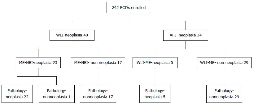
Figure 1 Flow diagram of neoplastic lesions detected by trimodal imaging endoscopy.
EGD: Esophagogastroduodenoscopy; WLI: White-light imaging; AFI: Autofluorescence imaging; ME: Magnifying endoscopy; NBI: Narrow-band imaging.
- Citation: Imaeda H, Hosoe N, Kashiwagi K, Ida Y, Nakamura R, Suzuki H, Saito Y, Yahagi N, Iwao Y, Kitagawa Y, Hibi T, Ogata H, Kanai T. Surveillance using trimodal imaging endoscopy after endoscopic submucosal dissection for superficial gastric neoplasia. World J Gastroenterol 2014; 20(43): 16311-16317
- URL: https://www.wjgnet.com/1007-9327/full/v20/i43/16311.htm
- DOI: https://dx.doi.org/10.3748/wjg.v20.i43.16311


