Copyright
©2014 Baishideng Publishing Group Inc.
World J Gastroenterol. Nov 7, 2014; 20(41): 15454-15461
Published online Nov 7, 2014. doi: 10.3748/wjg.v20.i41.15454
Published online Nov 7, 2014. doi: 10.3748/wjg.v20.i41.15454
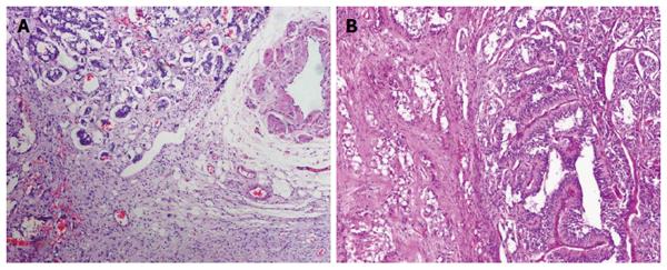
Figure 4 Photomicrographs of the pelvic cavity lesion.
A: At low-power fields, the lesion appeared to have an infiltrative margin without necrosis, hemorrhage or invasion of blood vessels; B: At high-power fields, the lesion exhibited an admixture of three types of tumor cells, and epithelioid cells were observed to be arranged in pseudoglandular structures (A: HE staining with original magnification × 100; B: HE staining with original magnification × 400). HE: Hematoxylin and eosin.
- Citation: Li B, Li Y, Tian XY, Luo BN, Li Z. Malignant gangliocytic paraganglioma of the duodenum with distant metastases and a lethal course. World J Gastroenterol 2014; 20(41): 15454-15461
- URL: https://www.wjgnet.com/1007-9327/full/v20/i41/15454.htm
- DOI: https://dx.doi.org/10.3748/wjg.v20.i41.15454


