Copyright
©2014 Baishideng Publishing Group Inc.
World J Gastroenterol. Oct 21, 2014; 20(39): 14442-14449
Published online Oct 21, 2014. doi: 10.3748/wjg.v20.i39.14442
Published online Oct 21, 2014. doi: 10.3748/wjg.v20.i39.14442
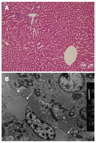
Figure 3 Histologic findings in the portal and central area from a rabbit in group 2.
A: Light microscopic finding shows no specific abnormality in the hepatic artery, hepatocytes, sinusoids or central hepatic veins (HE, × 200); B: Electron microscopic finding shows multiple fat vacuoles (arrows) within the vascular lumen. However, there is no evidence of interstitial edema. The integrity of the endothelium is well preserved.
- Citation: Kim YW, Park YM, Yoon S, Kim HJ, Park DY, Cho BM, Choi SH. Effect of intra-arterial infusion with triolein emulsion on rabbit liver. World J Gastroenterol 2014; 20(39): 14442-14449
- URL: https://www.wjgnet.com/1007-9327/full/v20/i39/14442.htm
- DOI: https://dx.doi.org/10.3748/wjg.v20.i39.14442


