Copyright
©2014 Baishideng Publishing Group Inc.
World J Gastroenterol. Aug 28, 2014; 20(32): 11160-11181
Published online Aug 28, 2014. doi: 10.3748/wjg.v20.i32.11160
Published online Aug 28, 2014. doi: 10.3748/wjg.v20.i32.11160
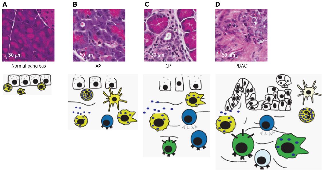
Figure 6 Immune cells in progressive pancreatic disease.
A: The normal pancreas contains sparse, mostly innate, inflammatory cells and lacks the dense stroma typically seen in chronic pancreatitis (CP) and pancreatic ductal adenocarcinoma (PDAC); B: Acute pancreatitis (AP) is characterized by acinar degranulation, edema and recruitment of mostly innate inflammatory cells, but also some T-lymphocytes in response to acinar damage. Pancreatic mast cells begin to degranulate; C: CP is characterized by development of stroma surrounding degranulated acinar cells, acinar-to-ductal metaplasia and edema. Additionally, there is an increased presence of macrophages, and T- and B-lymphocytes, and further degranulation of mast cells; D: Development of pancreatic intraepithelial neoplasias and subsequent PDAC leads to a significant increase in immunosuppressive cell types including tumor-associated macrophages, myeloid-derived suppressor cells, and Tregs. Degranulated mast cells, neutrophils, dendritic cells and B- and T-lymphocytes are also present. However, T-lymphocytes and dendritic cells are typically inhibited and defective, respectively. Size of immune cell represents relative abundance. Cell color denotes immune cell type: yellow, innate immune cells; blue, adaptive immune cells; green, immunosuppressive cells. For description of graphical representation of cell types, see Figure 3 legend. Cells that are inactivated or defective are represented by a lighter color.
- Citation: Inman KS, Francis AA, Murray NR. Complex role for the immune system in initiation and progression of pancreatic cancer. World J Gastroenterol 2014; 20(32): 11160-11181
- URL: https://www.wjgnet.com/1007-9327/full/v20/i32/11160.htm
- DOI: https://dx.doi.org/10.3748/wjg.v20.i32.11160


