Published online Aug 28, 2014. doi: 10.3748/wjg.v20.i32.11456
Revised: March 18, 2014
Accepted: May 12, 2014
Published online: August 28, 2014
Accidentally ingested foreign bodies, for the most part, pass through the gastrointestinal tract, but can cause several complications. Perforation is rare, but can occur in any segment of the gastrointestinal tract. Intestinal perforations due to foreign bodies are rarely diagnosed preoperatively as clinical symptoms are non-specific and they can mimic other abdominal conditions. We describe a case of a 48-year-old patient who was admitted to the emergency room because of severe abdominal pain of 5 d duration. A computed tomography scan showed an undefined liquid collection involving a linear image 35 mm in size, suggestive of a foreign body. On laparotomy, an abscess containing a fish bone was resected. As fish bone ingestion is usually not remembered by the patient, the diagnosis can be delayed. The preoperative diagnosis is frequently acute abdomen of unknown cause. A low threshold of suspicion along with a good clinical history and radiological studies are extremely important in order to make a correct diagnosis.
Core tip: Perforations due to fish bones are rare and have nonspecific symptoms, mimicking other abdominal conditions. A patient attended the emergency room due to severe abdominal pain of 5 d duration. A computed tomography scan showed an undefined liquid collection involving a linear image 35 mm in size. On laparotomy, an abscess containing a fish bone was resected.
- Citation: Wu CX, Wu BQ, Duan YF, Sun DL, Jiang Y. Rare case of omentum-wrapped abscess caused by a fish bone penetrating the terminal ileum. World J Gastroenterol 2014; 20(32): 11456-11459
- URL: https://www.wjgnet.com/1007-9327/full/v20/i32/11456.htm
- DOI: https://dx.doi.org/10.3748/wjg.v20.i32.11456
Foreign body ingestion is frequent and can cause several complications, perforation being the most frequent[1-3]. Perforation due to ingested foreign bodies in the adult population is most common secondary to accidental ingestion and is frequently caused by dietary foreign bodies, especially fish bones. Foreign bodies usually pass through the gastrointestinal tract without problems once beyond the esophagus[3,4]. Perforation occurs in about 1% of all foreign bodies ingested usually due to long and sharp objects such as fish bones, toothpicks, chicken bones and needles[3,4]. We herein report the diagnosis and treatment of a patient with omentum-wrapped abscess caused by a fish bone penetrating the terminal ileum.
A 48-year-old male patient attended the emergency room due to severe abdominal pain, mainly in the middle quadrants, of 5 d duration, without nausea or vomiting. Gastrointestinal transit was normal, without bleeding per rectum. There was no history of anorexia or weight loss. Respiratory and urinary symptoms were absent. On physical examination, the patient had generalized tenderness of the abdomen, which was maximal in the middle upper quadrant, signs of peritoneal inflammation with guarding, rebound, and tap tenderness, without Rovsing’s or Murphy’s sign, and peristaltic sounds were not audible. He was tachycardic with a fever of 39.6 °C. White blood cells count was 11.96 × 10⁹/μL, no anemia was found, and hepatic and pancreatic tests were normal.
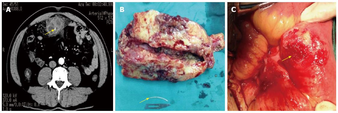
Abdominal ultrasound showed peritoneal effusion, a normal gallbladder, and the ileo-cecal appendix was not visualized. An abdominal computed tomography (CT) scan was performed, and in addition to moderate peritoneal effusion, an undefined liquid collection involving a linear image 35 mm in size was evident in the omental abscess, suggestive of a foreign body (Figure 1A).
When asked about his diet, the patient mentioned that some days earlier he had eaten fish, however, foreign body ingestion was not remembered. On this basis, a diagnosis of bowel perforation due to a foreign body (fish bone) was made.
Systemic antibiotics were initiated, and a median laparotomy was performed, revealing a purulent peritoneal effusion and an abscess in the greater omentum adjacent to the transverse colon. A fibrotic closed fistula between the terminal ileum and the abscess was found (Figure 1C), which was sectioned, and the abscess was resected en-bloc. There was no evident colon defect, and we performed a purse-string suture in the ileum side of the fibrotic fistula and an omental patch was used to cover it. Peritoneal lavage was carried out using saline solution and a drain was placed in the abdominal cavity. When the abscess was opened, a fish bone of approximately 35 mm was found (Figure 1B).
No complications occurred after surgery, the drain was removed on the fourth post-operative day (drainage was always serous), and the patient was discharged free of symptoms seven days after surgery. During the follow-up period of 3 mo, no sequelae were observed.
Perforation can occur in any segment of the gastrointestinal tract[2-4], however, the most common sites of perforation are in the distal ileum[5-7], the cecum and the left colon[8] due to their great angulation[3,4]. Most perforations occur in the straits and the angles of the gastrointestinal tract[9]. Ingestion of foreign bodies, although a fairly common problem in pediatrics, is relatively rare in adults and is mainly found in individuals with psychiatric disorders such as bipolar disorder, depression, or post-traumatic stress disorder[10]. Alcoholism, psychiatric illness, age extremes and the use of dentures are risk factors for foreign body ingestion[3,4]. Intestinal perforation by fish bones is rare, however, their ingestion is common. The risk of perforation is related to the length and shape of the object ingested[11]. The average time from ingestion of the foreign body to perforation is 10.4 d[12]. Clinical presentation can vary with acute or chronic symptoms[2,3]. Bowel perforation by foreign bodies can mimic other abdominal conditions such as acute appendicitis, acute diverticulitis, and perforated peptic ulcer[13]. As the patient usually does not remember fish bone ingestion, diagnosis can be delayed, with months between ingestion and perforation[1,3,4]. Fish bones located in a narrow segment of the bowel can erode the mucosa, causing bacterial dissemination. As this pathological process continues, perforation and an extramural abscess occur, which leads to acute abdominal pain[3]. In the case presented here, the patient had eaten fish some days before and fish bone ingestion was not remembered, however, as a fibrotic closed fistula was found, the ingestion causing perforation was thought to have occurred earlier.
X-rays can be used to detect foreign bodies. Plain radiography is helpful in locating metallic foreign bodies and pneumoperitoneum. As the perforation hole is small and normally covered with fibrin and omentum, pneumoperitoneum is rare, being present in only 20% of patients[3,4]. CT scanning is the most accurate exam with fish bones appearing like linear images with calcic density inside an inflamed area[3]. However, CT scanning has some weaknesses: lack of awareness by the radiologist if there is no clinical suspicion; the use of oral and/or intravenous contrast can make it difficult to visualize fish bones[3]. Colon perforation can have the same radiologic and pathologic characteristics as intestinal inflammatory disease[14]. CT scanning was used in our patient to aid the diagnosis and showed a linear image inside a liquid collection suggestive of a foreign body, without pneumoperitoneum.
Treatment depends on the age of the patient and symptoms, the nature and type of foreign body and anatomical location, especially if impacted. The management may consist of conservative or interventional methods, endoscopic, laparoscopic or open surgery. Surgery is the treatment of choice for bowel perforation, and is most commonly performed by laparotomy due to its advantages in localizing the perforation, closure or repair of the defect, and peritoneal lavage. However, laparoscopy has been reported in some studies to be as good as laparotomy[15]. In our case, laparotomy was chosen, and the abscess adjacent to the transverse colon was resected en-bloc to avoid spreading pus into the abdominal cavity, and peritoneal lavage was performed to clean the purulent effusion. No defect was found in the terminal ileum, as the fistula was fibrotic and occluded, this may have been because there was a long time between perforation and surgery, with the greater omentum tapering the defect. Intestinal perforation treatment usually involves bowel resection, however, the most common treatment is suture of the perforation[8]. For this reason, the closed fistula was sectioned and a purse-string suture was performed and covered with an omental patch for safety. The patient was discharged free of symptoms on the 7th post-operative day, which is consistent with other similar case reports.
A 48-year-old patient attended the emergency room due to severe abdominal pain of 5 d duration.
On laparotomy, an abscess containing a fish bone was resected.
Bowel perforation by foreign bodies can mimic other abdominal conditions such as acute appendicitis, acute diverticulitis, and perforated peptic ulcer.
The patient was tachycardic with a fever of 39.6 °C. Blood samples revealed a white blood cells of 11.96 × 10⁹/μL, no anemia, and normal hepatic and pancreatic tests.
A computed tomography scan showed an undefined liquid collection involving a linear image 35 mm in size, suggestive of a foreign body.
Fish bone, fat and fibrous tissue with chronic suppurative inflammation, multifocal abscess formation, and histiocytosis.
Laparotomy was performed and the abscess was resected en-bloc to avoid spreading pus into the abdominal cavity.
A low threshold of suspicion along with a good clinical history and radiological studies are extremely important in order to make a correct diagnosis.
The authors reported a patient with omentum-wrapped abscess caused by a fish bone penetrating the terminal ileum, and presented the imaging and surgical findings of the location of the terminal ileum perforation, the size of the abscess and the length of the fish bone in the abscess.
P- Reviewer: Faenza S, Skok P S- Editor: Gou SX L- Editor: A E- Editor: Zhang DN
| 1. | Puia IC, Puia VR, Andreescu A, Cristea PG. [Ascending colon perforation by ingested fruit stones]. Chirurgia (Bucur). 2011;106:825-827. [PubMed] [Cited in This Article: ] |
| 2. | Chiu JJ, Chen TL, Zhan YL. Perforation of the transverse colon by a fish bone: a case report. J Emerg Med. 2009;36:345-347. [PubMed] [DOI] [Cited in This Article: ] [Cited by in Crossref: 14] [Cited by in F6Publishing: 15] [Article Influence: 0.9] [Reference Citation Analysis (0)] |
| 3. | Sierra-Solís A. [Bowel perforations due to fish bones: rare and curious]. Semergen. 2013;39:117-118. [PubMed] [DOI] [Cited in This Article: ] [Cited by in Crossref: 4] [Cited by in F6Publishing: 4] [Article Influence: 0.3] [Reference Citation Analysis (0)] |
| 4. | Joglekar S, Rajput I, Kamat S, Downey S. Sigmoid perforation caused by an ingested chicken bone presenting as right iliac fossa pain mimicking appendicitis: a case report. J Med Case Rep. 2009;3:7385. [PubMed] [DOI] [Cited in This Article: ] [Cited by in Crossref: 15] [Cited by in F6Publishing: 17] [Article Influence: 1.1] [Reference Citation Analysis (0)] |
| 5. | Eisen GM, Baron TH, Dominitz JA, Faigel DO, Goldstein JL, Johanson JF, Mallery JS, Raddawi HM, Vargo JJ, Waring JP. Guideline for the management of ingested foreign bodies. Gastrointest Endosc. 2002;55:802-806. [PubMed] [Cited in This Article: ] |
| 6. | Murshid KR, Khairy GE. Laparoscopic removal of a foreign body from the intestine. J R Coll Surg Edinb. 1998;43:109-111. [PubMed] [Cited in This Article: ] |
| 7. | Goh BK, Chow PK, Quah HM, Ong HS, Eu KW, Ooi LL, Wong WK. Perforation of the gastrointestinal tract secondary to ingestion of foreign bodies. World J Surg. 2006;30:372-377. [PubMed] [Cited in This Article: ] |
| 8. | Pinero Madrona A, Fernández Hernández JA, Carrasco Prats M, Riquelme Riquelme J, Parrila Paricio P. Intestinal perforation by foreign bodies. Eur J Surg. 2000;166:307-309. [PubMed] [Cited in This Article: ] |
| 9. | Singh RP, Gardner JA. Perforation of the sigmoid colon by swallowed chicken bone: case reports and review of literature. Int Surg. 1981;66:181-183. [PubMed] [Cited in This Article: ] |
| 10. | Mesina C, Vasile I, Valcea DI, Pasalega M, Calota F, Paranescu H, Dumitrescu T, Mirea C, Mogoanta S. Problems of diagnosis and treatment caused by ingested foreign bodies. Chirurgia (Bucur). 2013;108:400-406. [PubMed] [Cited in This Article: ] |
| 11. | Sarliève P, Delabrousse E, Michalakis D, Robert A, Guichard G, Kastler B. Multidetector ct diagnosis of jejunal perforation by a chicken bone. JBR-BTR. 2004;87:294-295. [PubMed] [Cited in This Article: ] |
| 12. | Rodríguez-Hermosa JI, Codina-Cazador A, Sirvent JM, Martín A, Gironès J, Garsot E. Surgically treated perforations of the gastrointestinal tract caused by ingested foreign bodies. Colorectal Dis. 2008;10:701-707. [PubMed] [Cited in This Article: ] |
| 13. | Yao CC, Yang CC, Liew SC, Lin CS. Small bowel perforation caused by a sharp bone: laparoscopic diagnosis and treatment. Surg Laparosc Endosc Percutan Tech. 1999;9:226-227. [PubMed] [Cited in This Article: ] |
| 14. | Ali FE, Al-Busairi WA, Esbaita EY, Al-Bustan MA. Chronic perforation of the sigmoid colon by foreign body. Curr Surg. 2005;62:419-422. [PubMed] [Cited in This Article: ] |
| 15. | Arora S, Ashrafian H, Smock ED, Ng P. Total laparoscopic repair of sigmoid foreign body perforation. J Laparoendosc Adv Surg Tech A. 2009;19:401-403. [PubMed] [DOI] [Cited in This Article: ] [Cited by in Crossref: 10] [Cited by in F6Publishing: 12] [Article Influence: 0.8] [Reference Citation Analysis (0)] |


