Copyright
©2014 Baishideng Publishing Group Inc.
World J Gastroenterol. Aug 28, 2014; 20(32): 11456-11459
Published online Aug 28, 2014. doi: 10.3748/wjg.v20.i32.11456
Published online Aug 28, 2014. doi: 10.3748/wjg.v20.i32.11456
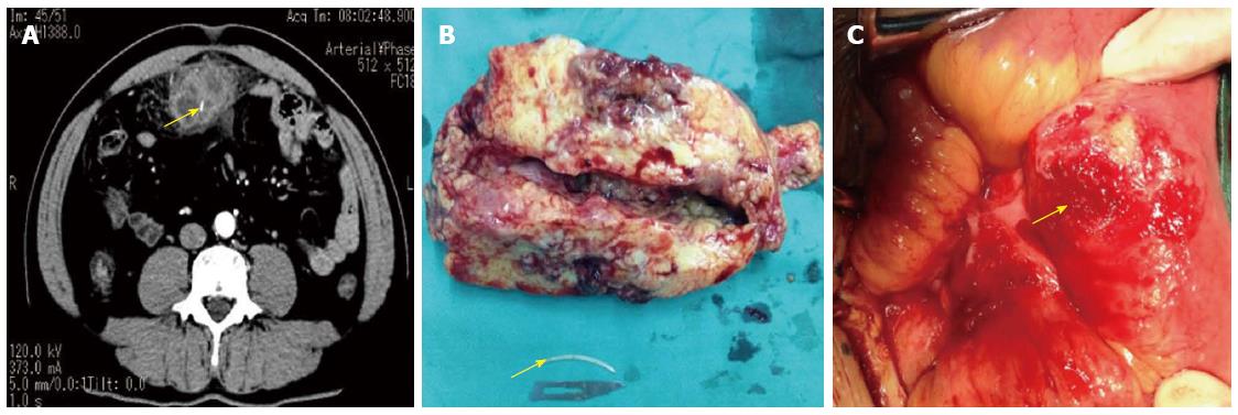
Figure 1 Computed tomography, resected abscess and fish bone and the lacation of perforation in terminal ileum.
A: Axial and sagittal slides of abdominal computed tomography scan. It is visible an undefined liquid collection involving a linear image suggestive of foreign body (arrow); B: The resected abscess size and fishbone (arrow); C: The perforation in terminal ileum (arrow).
- Citation: Wu CX, Wu BQ, Duan YF, Sun DL, Jiang Y. Rare case of omentum-wrapped abscess caused by a fish bone penetrating the terminal ileum. World J Gastroenterol 2014; 20(32): 11456-11459
- URL: https://www.wjgnet.com/1007-9327/full/v20/i32/11456.htm
- DOI: https://dx.doi.org/10.3748/wjg.v20.i32.11456


