Copyright
©2014 Baishideng Publishing Group Co.
World J Gastroenterol. Jan 21, 2014; 20(3): 857-862
Published online Jan 21, 2014. doi: 10.3748/wjg.v20.i3.857
Published online Jan 21, 2014. doi: 10.3748/wjg.v20.i3.857
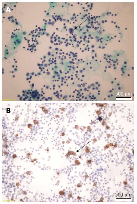
Figure 2 Cytological analysis of the ascitic fluid.
A: Papanicolaou staining showed a few large immunoblastic-like atypical cells with large nuclei and prominent nucleoli (arrow). Magnification: × 400; B: Immunohistochemistry staining showed large, CD20-positive lymphoid cells (arrow). Magnification: × 400.
- Citation: Nussinson E, Shibli F, Shahbari A, Rock W, Elias M, Elmalah I. Primary effusion lymphoma-like lymphoma in a patient with inflammatory bowel disease. World J Gastroenterol 2014; 20(3): 857-862
- URL: https://www.wjgnet.com/1007-9327/full/v20/i3/857.htm
- DOI: https://dx.doi.org/10.3748/wjg.v20.i3.857


