Copyright
©2014 Baishideng Publishing Group Inc.
World J Gastroenterol. Jul 28, 2014; 20(28): 9621-9625
Published online Jul 28, 2014. doi: 10.3748/wjg.v20.i28.9621
Published online Jul 28, 2014. doi: 10.3748/wjg.v20.i28.9621
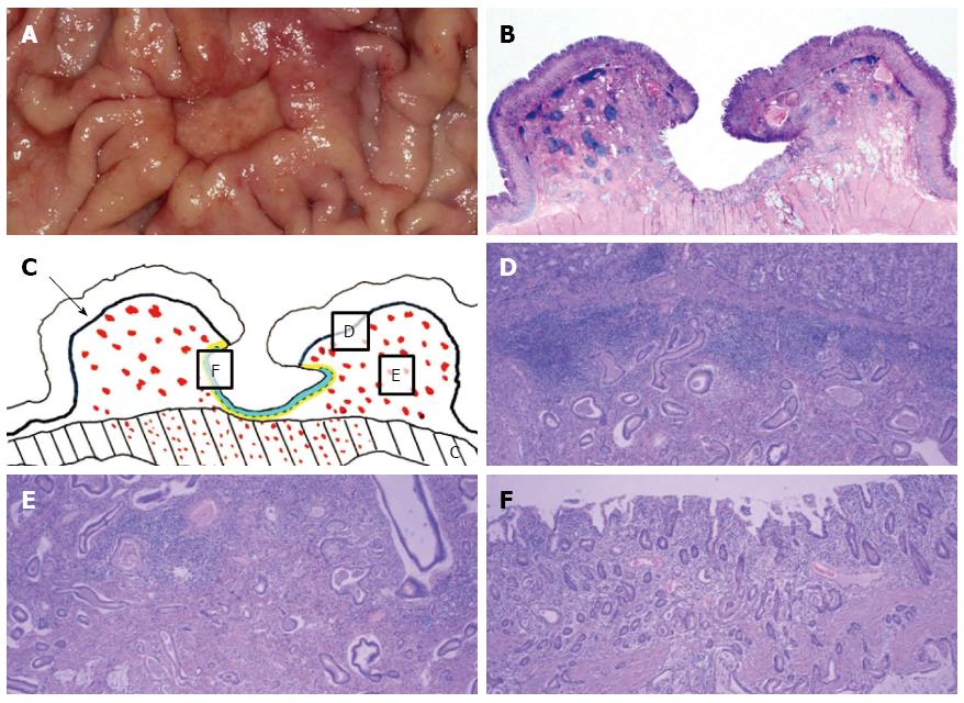
Figure 3 Gross surgical specimen and microscopic findings with reconstruction mapping.
A: Gross specimen. A deep round ulcerofungating lesion among the gastric folds with focal fold conversions; B: Gross histopathologic finding for a coronal section (hematoxylin and eosin stain, × 1). The large ulcer was covered with superficial regenerative epithelium, and bunches of cancer cells were scattered below the epithelium; C: Reconstruction mapping. The four solid lines separate the mucosa, submucosa, muscularis propria and serosa. The thick solid line is the muscularis mucosa layer (arrow). The red dots indicate cancer cells. The dotted line is an imaginary line that separates regenerative epithelium from the submucosal layer. Above this line, the light blue area is a regenerative epithelial area. Underneath, there is yellow area in which no cancer cells are found; D-F: Each figure corresponds with a part of Figure 3C (hematoxylin and eosin stain, × 40). Multiple cancer cells were noted in the submucosa, muscle and serosa. However, there were no cancer cells in the mucosal layer in the ulcer, including at the margins and base of the ulcer.
- Citation: Hur J, Chang JH, Kim BK, Ko HY, Lee JH, Kim SJ, Song MA, Kim TH, Kim CW, Han SW. Undiagnosed Borrmann type II gastric cancer due to necrosis and regenerative epithelium. World J Gastroenterol 2014; 20(28): 9621-9625
- URL: https://www.wjgnet.com/1007-9327/full/v20/i28/9621.htm
- DOI: https://dx.doi.org/10.3748/wjg.v20.i28.9621


