Copyright
©2014 Baishideng Publishing Group Inc.
World J Gastroenterol. Jul 14, 2014; 20(26): 8745-8750
Published online Jul 14, 2014. doi: 10.3748/wjg.v20.i26.8745
Published online Jul 14, 2014. doi: 10.3748/wjg.v20.i26.8745
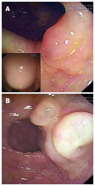
Figure 1 Endoscopic findings for the two cases.
A round, semi-transparent and wide-based submucosal lesion at the ascending colon in Case 1 (A) and a sharply marginated, lobular submucosal lesion at the sigmoid colon in Case 2 (B) with gentle slopes and smooth surfaces are noted. Left bottom of Figure 1A shows that when the patient's position was altered, the shape of the mass changed and was fluctuant on palpation.
- Citation: Zhuo CH, Shi DB, Ying MG, Cheng YF, Wang YW, Zhang WM, Cai SJ, Li XX. Laparoscopic segmental colectomy for colonic lymphangiomas: A definitive, minimally invasive surgical option. World J Gastroenterol 2014; 20(26): 8745-8750
- URL: https://www.wjgnet.com/1007-9327/full/v20/i26/8745.htm
- DOI: https://dx.doi.org/10.3748/wjg.v20.i26.8745


