Copyright
©2014 Baishideng Publishing Group Co.
World J Gastroenterol. Jan 14, 2014; 20(2): 384-400
Published online Jan 14, 2014. doi: 10.3748/wjg.v20.i2.384
Published online Jan 14, 2014. doi: 10.3748/wjg.v20.i2.384
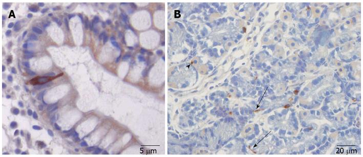
Figure 2 The gut endocrine cells.
A: A chromogranin-A-immunoreactive endocrine cell in the ileum. The endocrine cell extends from the basal membrane of the mucosa that project into the gut lumen; B: Somatostatin-immunoreactive cells in the gastric oxyntic mucosa. Note the long cytoplasmic processes (arrows), which can occasionally be seen to end at the base of parietal cells (arrowhead).
- Citation: El-Salhy M, Gundersen D, Gilja OH, Hatlebakk JG, Hausken T. Is irritable bowel syndrome an organic disorder? World J Gastroenterol 2014; 20(2): 384-400
- URL: https://www.wjgnet.com/1007-9327/full/v20/i2/384.htm
- DOI: https://dx.doi.org/10.3748/wjg.v20.i2.384


