Copyright
©2014 Baishideng Publishing Group Co.
World J Gastroenterol. May 14, 2014; 20(18): 5375-5388
Published online May 14, 2014. doi: 10.3748/wjg.v20.i18.5375
Published online May 14, 2014. doi: 10.3748/wjg.v20.i18.5375
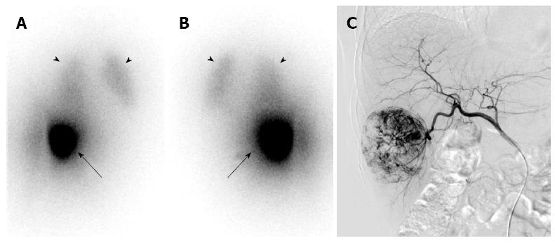
Figure 3 A Bremsstrahlung scan of 90Y-microsphere transarterial radioembolization.
Anterior (A) and posterior (B) planar scan images show hot uptake (arrows) in the right lobe of the liver, which is well correlated with findings on angiography (C), in spite of relatively poor image quality with blurring. Some liver-to-lung shunt activities are shown in the lungs (arrowheads).
- Citation: Eo JS, Paeng JC, Lee DS. Nuclear imaging for functional evaluation and theragnosis in liver malignancy and transplantation. World J Gastroenterol 2014; 20(18): 5375-5388
- URL: https://www.wjgnet.com/1007-9327/full/v20/i18/5375.htm
- DOI: https://dx.doi.org/10.3748/wjg.v20.i18.5375


