Copyright
©2014 Baishideng Publishing Group Co.
World J Gastroenterol. Apr 21, 2014; 20(15): 4446-4452
Published online Apr 21, 2014. doi: 10.3748/wjg.v20.i15.4446
Published online Apr 21, 2014. doi: 10.3748/wjg.v20.i15.4446
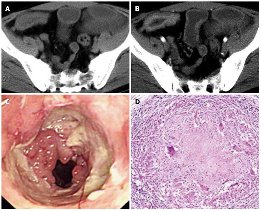
Figure 1 Computed tomography, endoscopic and pathological changes of intestinal tuberculosis in a 38-year-old man.
A: Plain computed tomography (CT) scan showed bowel-wall thickening (7.2 mm) and intestinal stricture in the ileocecum; B: During the arterial phase, contrast-enhanced CT demonstrated moderately stratified enhancement; C: Endoscopic examination showed rodent-like ulcer, ring-like ulcer and intestinal stricture in the ileocecum; D: Microscopic findings showed granulomas with caseous necrosis (hematoxylin and eosin staining; original magnification, 400 ×).
- Citation: Zhu QQ, Zhu WR, Wu JT, Chen WX, Wang SA. Comparative study of intestinal tuberculosis and primary small intestinal lymphoma. World J Gastroenterol 2014; 20(15): 4446-4452
- URL: https://www.wjgnet.com/1007-9327/full/v20/i15/4446.htm
- DOI: https://dx.doi.org/10.3748/wjg.v20.i15.4446


