Copyright
©2013 Baishideng Publishing Group Co.
World J Gastroenterol. Mar 7, 2013; 19(9): 1396-1404
Published online Mar 7, 2013. doi: 10.3748/wjg.v19.i9.1396
Published online Mar 7, 2013. doi: 10.3748/wjg.v19.i9.1396
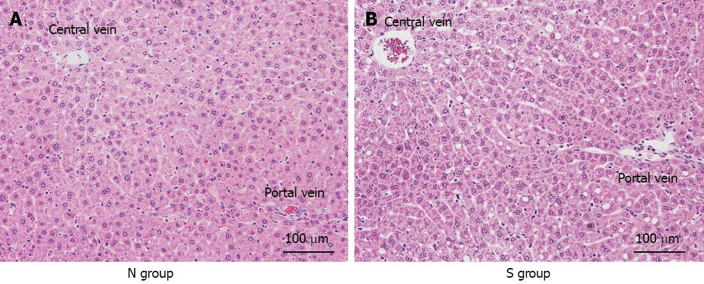
Figure 7 Histological findings after 120 min of reperfusion.
In the normal liver (N) group, hepatocyte structure was strongly impaired compared with the mild steatotic liver (S) group, and sinusoidal narrowing was observed.
- Citation: Ogawa K, Kondo T, Tamura T, Matsumura H, Fukunaga K, Oda T, Ohkohchi N. Influence of Kupffer cells and platelets on ischemia-reperfusion injury in mild steatotic liver. World J Gastroenterol 2013; 19(9): 1396-1404
- URL: https://www.wjgnet.com/1007-9327/full/v19/i9/1396.htm
- DOI: https://dx.doi.org/10.3748/wjg.v19.i9.1396


