Copyright
©2013 Baishideng Publishing Group Co.
World J Gastroenterol. Mar 7, 2013; 19(9): 1396-1404
Published online Mar 7, 2013. doi: 10.3748/wjg.v19.i9.1396
Published online Mar 7, 2013. doi: 10.3748/wjg.v19.i9.1396
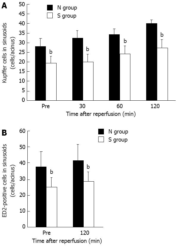
Figure 4 The number of Kupffer cells in sinusoids (mean ± SD).
A:The number of Kupffer cells in sinusoids observed in the intravital microscopy system was decreased significantly in the mild steatotic liver (S) group compared to the normal liver (N) group at any point in time before and after ischemia reperfusion. n = 6. bP < 0.01 vs the N group; B: In immunohistochemical staining, the number of ED2-positive cells was significantly less in the S group than in the N group. n = 6. bP < 0.01 vs the N group.
- Citation: Ogawa K, Kondo T, Tamura T, Matsumura H, Fukunaga K, Oda T, Ohkohchi N. Influence of Kupffer cells and platelets on ischemia-reperfusion injury in mild steatotic liver. World J Gastroenterol 2013; 19(9): 1396-1404
- URL: https://www.wjgnet.com/1007-9327/full/v19/i9/1396.htm
- DOI: https://dx.doi.org/10.3748/wjg.v19.i9.1396


