Copyright
©2013 Baishideng Publishing Group Co.
World J Gastroenterol. Feb 14, 2013; 19(6): 855-865
Published online Feb 14, 2013. doi: 10.3748/wjg.v19.i6.855
Published online Feb 14, 2013. doi: 10.3748/wjg.v19.i6.855
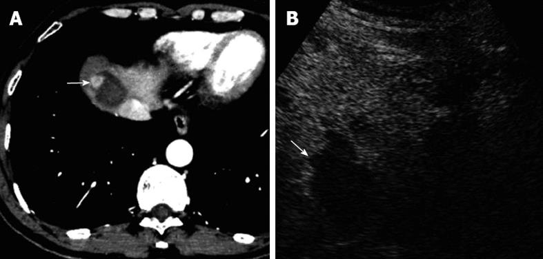
Figure 5 A 54-year-old male patient with hepatocellular carcinoma.
Three months after radiofrequency ablation in combination with ethanol ablation for hepatocellular carcinoma in segment 8 of the liver. A: Contrast-enhanced computed tomography showed local tumor progression (arrow) at the periphery of the treated area; B: On contrast-enhanced ultrasound, local tumor progression (arrow) could not be detected, and the treated area was not clearly observed due to unfavorable location near the liver dome.
- Citation: Zheng SG, Xu HX, Lu MD, Xie XY, Xu ZF, Liu GJ, Liu LN. Role of contrast-enhanced ultrasound in follow-up assessment after ablation for hepatocellular carcinoma. World J Gastroenterol 2013; 19(6): 855-865
- URL: https://www.wjgnet.com/1007-9327/full/v19/i6/855.htm
- DOI: https://dx.doi.org/10.3748/wjg.v19.i6.855


