Copyright
©2013 Baishideng Publishing Group Co.
World J Gastroenterol. Sep 21, 2013; 19(35): 5940-5942
Published online Sep 21, 2013. doi: 10.3748/wjg.v19.i35.5940
Published online Sep 21, 2013. doi: 10.3748/wjg.v19.i35.5940
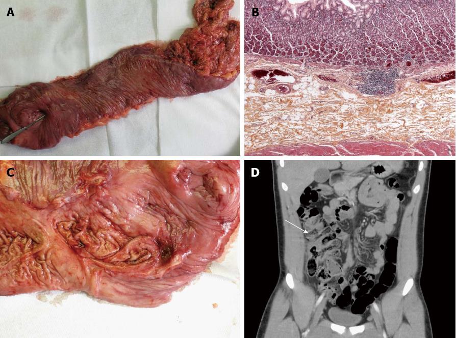
Figure 1 Colonic duplication.
A: Macroscopic analysis of the resected colonic duplication: colonic duplication with a pen showing the anastomosis with the right colon; B: Pathological analysis of the duplication: pathological analysis showing intestinal duplication with a smooth muscle coat and an ectopic gastric mucosal lining; C: Bleeding ulcer in the colonic duplication: macroscopic view of a large bleeding ulcer near the anastomosis between the duplicated and the right colon; D: Computed tomography scan coronal view of the colonic duplication: retrospective coronal reconstruction of the emergency computed tomography scan showing the duplicated right colon (arrow) which was not visible in the axial view.
- Citation: Jacques J, Projetti F, Legros R, Valgueblasse V, Sarabi M, Carrier P, Fredon F, Bouvier S, Loustaud-Ratti V, Sautereau D. Obscure bleeding colonic duplication responds to proton pump inhibitor therapy. World J Gastroenterol 2013; 19(35): 5940-5942
- URL: https://www.wjgnet.com/1007-9327/full/v19/i35/5940.htm
- DOI: https://dx.doi.org/10.3748/wjg.v19.i35.5940


