Copyright
©2013 Baishideng Publishing Group Co.
World J Gastroenterol. Sep 7, 2013; 19(33): 5464-5472
Published online Sep 7, 2013. doi: 10.3748/wjg.v19.i33.5464
Published online Sep 7, 2013. doi: 10.3748/wjg.v19.i33.5464
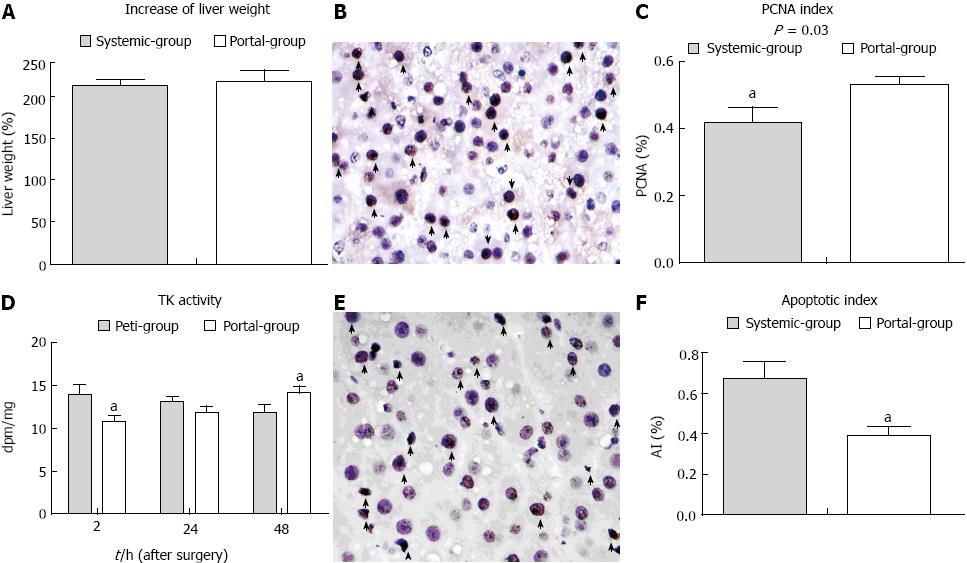
Figure 4 The rate of liver remnant regeneration was elevated and apoptosis was attenuated in the Portal group compared with the Systemic group.
A: The rate of increase of liver volume in the two groups; B: Proliferating cell nuclear antigen (PCNA) staining in the liver remnant (arrows indicate stained positive cells × 400 magnification). A large number of transferase dUTP nick end labeling (TUNEL)-positive cells (arrows) in the liver remnant (× 400 magnification); C: Microphotometric evaluation of the PCNA index in PCNA-stained tissue at 48 h PH; D: The change in thymidine kinase activity in the two groups; E: TUNEL staining at 48 h PH; F: Microphotometric evaluation of the apoptotic index (AI) in TUNEL-stained tissue at 48 h PH. aP < 0.05 indicates a significant difference between the groups.
- Citation: Hou P, Chen C, Tu YL, Zhu ZM, Tan JW. Extracorporeal continuous portal diversion plus temporal plasmapheresis for “small-for-size” syndrome. World J Gastroenterol 2013; 19(33): 5464-5472
- URL: https://www.wjgnet.com/1007-9327/full/v19/i33/5464.htm
- DOI: https://dx.doi.org/10.3748/wjg.v19.i33.5464


