Copyright
©2013 Baishideng Publishing Group Co.
World J Gastroenterol. Aug 14, 2013; 19(30): 5021-5024
Published online Aug 14, 2013. doi: 10.3748/wjg.v19.i30.5021
Published online Aug 14, 2013. doi: 10.3748/wjg.v19.i30.5021
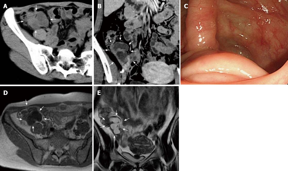
Figure 1 Preoperative image of the mucocele.
A: Computed tomography (CT) (axial image); a tumor was detected (arrows); B: CT (coronal image); a tumor was detected (arrows); C: Colonoscopy; submucosal tumor-like elevations of the appendiceal orifice; D: Magnetic resonance imaging (MRI) (T1-WI, axial image); E: MRI (T2-WI, coronal image).
- Citation: Tsuda M, Yamashita Y, Azuma S, Akamatsu T, Seta T, Urai S, Uenoyama Y, Deguchi Y, Ono K, Chiba T. Mucocele of the appendix due to endometriosis: A rare case report. World J Gastroenterol 2013; 19(30): 5021-5024
- URL: https://www.wjgnet.com/1007-9327/full/v19/i30/5021.htm
- DOI: https://dx.doi.org/10.3748/wjg.v19.i30.5021


