Copyright
©2013 Baishideng Publishing Group Co.
World J Gastroenterol. May 14, 2013; 19(18): 2826-2829
Published online May 14, 2013. doi: 10.3748/wjg.v19.i18.2826
Published online May 14, 2013. doi: 10.3748/wjg.v19.i18.2826
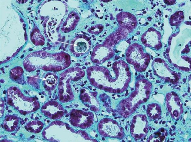
Figure 4 Renal biopsy showing acute tubular necrosis: Tubules dilated with sometimes a low-lying epithelium and pleomorphic nuclei.
The brush border of the proximal tubular cells is missing. The lumen of some tubules contains rare tubular cell necrosis (white arrows). There is a diffuse interstitial edema (Masson’s trichrome, ×40).
- Citation: Dupré A, Gagnière J, Tixier L, Ines DD, Perbet S, Pezet D, Buc E. Massive hepatic necrosis with toxic liver syndrome following portal vein ligation. World J Gastroenterol 2013; 19(18): 2826-2829
- URL: https://www.wjgnet.com/1007-9327/full/v19/i18/2826.htm
- DOI: https://dx.doi.org/10.3748/wjg.v19.i18.2826


