Copyright
©2013 Baishideng Publishing Group Co.
World J Gastroenterol. May 14, 2013; 19(18): 2826-2829
Published online May 14, 2013. doi: 10.3748/wjg.v19.i18.2826
Published online May 14, 2013. doi: 10.3748/wjg.v19.i18.2826
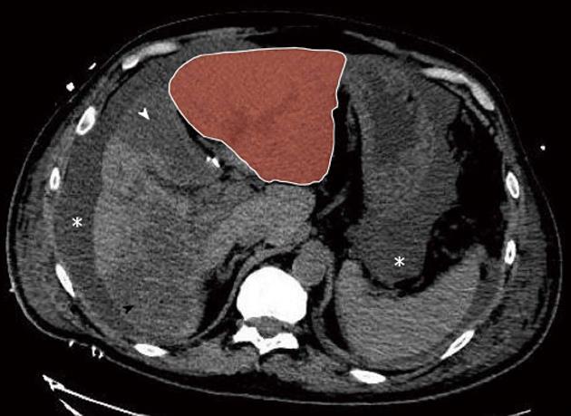
Figure 3 Postoperative day 2 non-enhanced multidetector computed tomograph scan.
Massive hepatic necrosis of the medial segment of the left lobe (segment 4, white arrowhead) and of a large part of the right lobe (black arrowhead) are shown, with remarkable hypertrophy of the lateral segments of the left lobe (red zone). Note also ascites (asterisk).
- Citation: Dupré A, Gagnière J, Tixier L, Ines DD, Perbet S, Pezet D, Buc E. Massive hepatic necrosis with toxic liver syndrome following portal vein ligation. World J Gastroenterol 2013; 19(18): 2826-2829
- URL: https://www.wjgnet.com/1007-9327/full/v19/i18/2826.htm
- DOI: https://dx.doi.org/10.3748/wjg.v19.i18.2826


