Copyright
©2013 Baishideng Publishing Group Co.
World J Gastroenterol. May 14, 2013; 19(18): 2826-2829
Published online May 14, 2013. doi: 10.3748/wjg.v19.i18.2826
Published online May 14, 2013. doi: 10.3748/wjg.v19.i18.2826
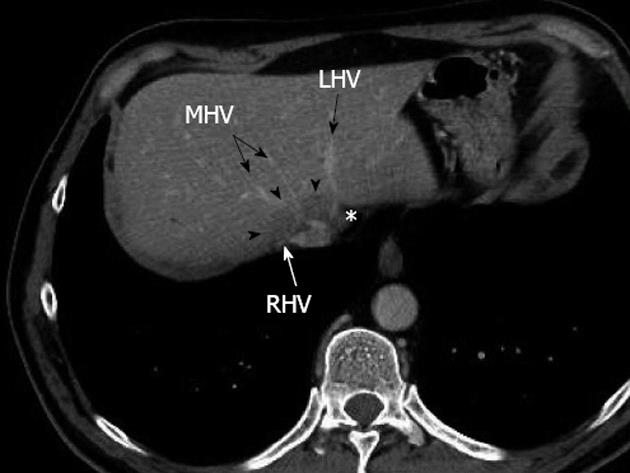
Figure 1 Preoperative multidetector computed tomograph at portal venous phase showing a large metastasis (arrowheads) encircling the confluence of the right hepatic vein and middle hepatic vein, without any thrombosis, and displacing the end of the left hepatic vein (asterisk).
RHV: Right hepatic vein; MHV: Middle hepatic vein; LHV: Left hepatic vein.
- Citation: Dupré A, Gagnière J, Tixier L, Ines DD, Perbet S, Pezet D, Buc E. Massive hepatic necrosis with toxic liver syndrome following portal vein ligation. World J Gastroenterol 2013; 19(18): 2826-2829
- URL: https://www.wjgnet.com/1007-9327/full/v19/i18/2826.htm
- DOI: https://dx.doi.org/10.3748/wjg.v19.i18.2826


