Copyright
©2013 Baishideng Publishing Group Co.
World J Gastroenterol. May 7, 2013; 19(17): 2638-2649
Published online May 7, 2013. doi: 10.3748/wjg.v19.i17.2638
Published online May 7, 2013. doi: 10.3748/wjg.v19.i17.2638
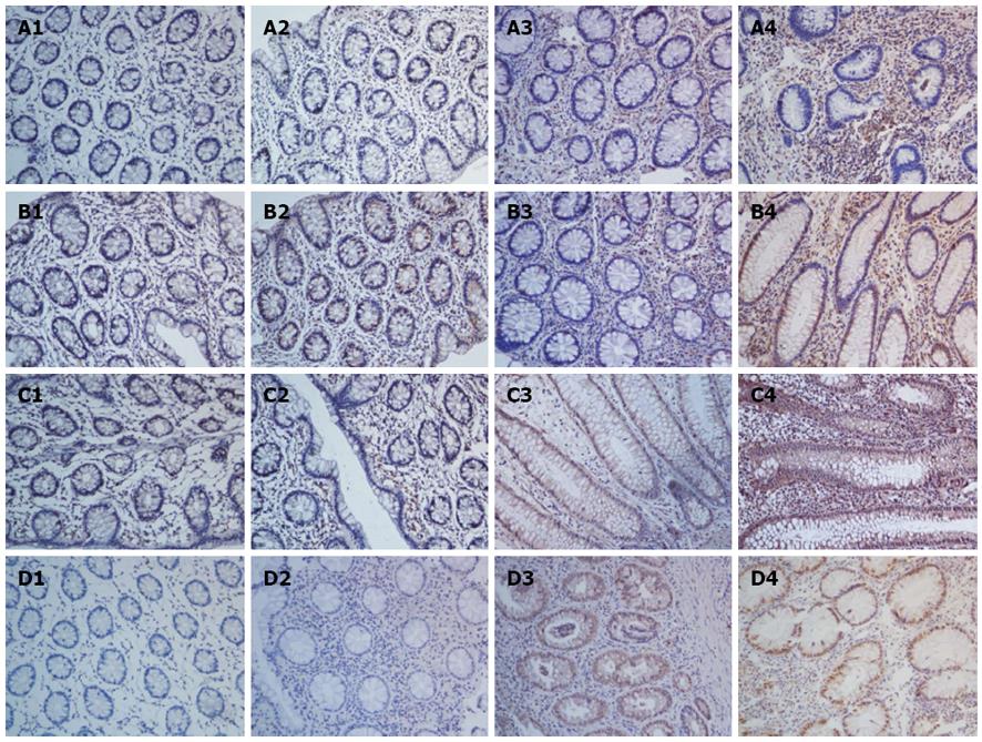
Figure 1 Expression and distribution of interleukin-22 and its related proteins in colonic biopsy specimens as analyzed by immunohistochemistry (× 200).
A1-A4: Expression and distribution of interleukin (IL)-22 in colonic biopsy specimens from the control, inactive ulcerative colitis (UC), mild-moderate UC, and severe UC tissues, respectively; B1-B4: The expression and distribution of IL-23 in colonic biopsy specimens from the control, inactive UC, mild-moderate UC, and severe UC tissues; C1-C4: The expression and distribution of IL-22R1 in colonic biopsy specimens from the control, inactive UC, mild-moderate UC, and severe UC tissues; D1-D4: The expression and distribution of p-STAT3 (S727) in colonic biopsy specimens from the control, inactive UC, mild-moderate UC, and severe UC tissues.
- Citation: Yu LZ, Wang HY, Yang SP, Yuan ZP, Xu FY, Sun C, Shi RH. Expression of interleukin-22/STAT3 signaling pathway in ulcerative colitis and related carcinogenesis. World J Gastroenterol 2013; 19(17): 2638-2649
- URL: https://www.wjgnet.com/1007-9327/full/v19/i17/2638.htm
- DOI: https://dx.doi.org/10.3748/wjg.v19.i17.2638


