Copyright
©2012 Baishideng Publishing Group Co.
World J Gastroenterol. Dec 14, 2012; 18(46): 6701-6708
Published online Dec 14, 2012. doi: 10.3748/wjg.v18.i46.6701
Published online Dec 14, 2012. doi: 10.3748/wjg.v18.i46.6701
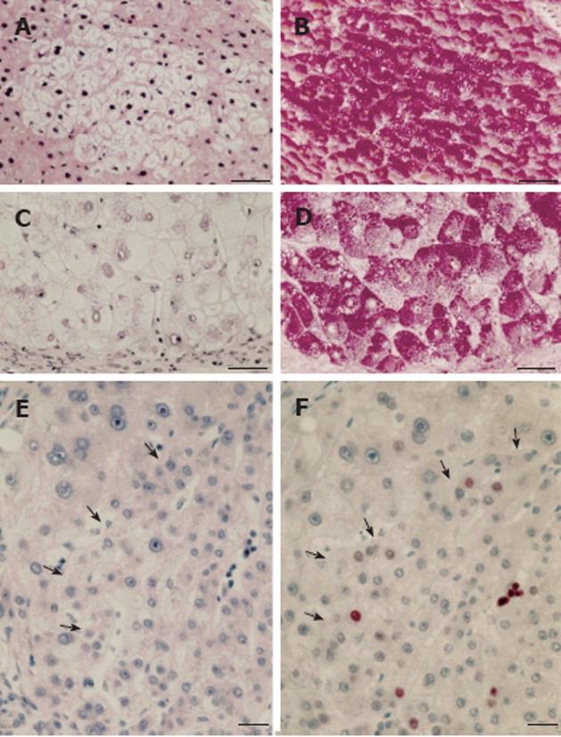
Figure 3 Human focal (A, B) and nodular (C, D) hepatic glycogenosis, and more advanced mixed cell populations (E, F) with intrafocal small cell change.
A, B: Hepatic vein. Perivenular glycogenolytic (clear cell) focus in an hepatocellular carcinoma (HCC)-bearing liver with hepatitis B virus (HBV)-associated cirrhosis, demonstrated in serial sections with hematoxylin and eosin (A) and periodic acid schiff (PAS) (B)-reaction counterstained with orange G and iron hematoxilin (Tri-PAS). Bar: 100 μm; C, D: Glycogenotic (clear cell) nodule in an HCC-free liver with HBV-associated cirrhosis demonstrated in serial sections with hematoxylin-eosin (C) and Tri-PAS (D). Bar: 100 μm; E, F: Hematoxylin and eosin (E), proliferating cell nuclear antigen (F) immunostaining. Increased cell proliferation (arrows) in a mixed cell focus with less pronounced glycogen storage (not shown) and with intrafocal small cell change in liver with cryptogenic cirrhosis, demonstrated in serial sections. Bar: 50 μm, from Su et al[68].
- Citation: Bannasch P. Glycogenotic hepatocellular carcinoma with glycogen-ground-glass hepatocytes: A heuristically highly relevant phenotype. World J Gastroenterol 2012; 18(46): 6701-6708
- URL: https://www.wjgnet.com/1007-9327/full/v18/i46/6701.htm
- DOI: https://dx.doi.org/10.3748/wjg.v18.i46.6701


