Copyright
©2012 Baishideng Publishing Group Co.
World J Gastroenterol. Nov 21, 2012; 18(43): 6216-6225
Published online Nov 21, 2012. doi: 10.3748/wjg.v18.i43.6216
Published online Nov 21, 2012. doi: 10.3748/wjg.v18.i43.6216
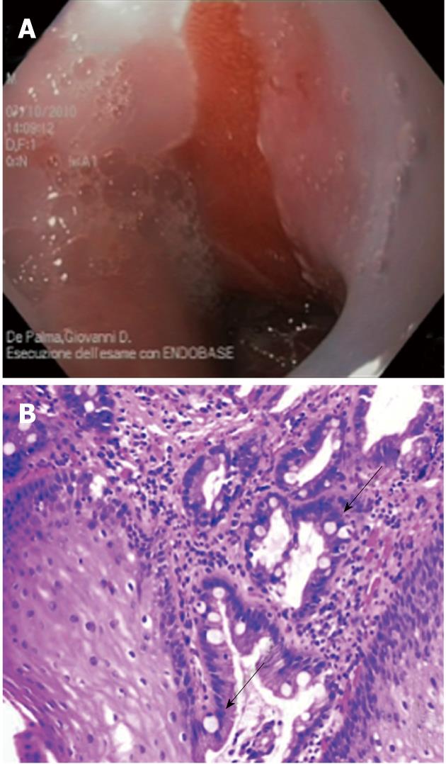
Figure 1 Endoscopic and histologic images of Barrett’s esophagus.
A: Endoscopic view of salmon-colored mucosa above the gastro-esophageal junction; B: Intestinal metaplasia with goblet cells (arrows) was found in biopsy specimens at histology.
- Citation: Palma GDD. Management strategies of Barrett's esophagus. World J Gastroenterol 2012; 18(43): 6216-6225
- URL: https://www.wjgnet.com/1007-9327/full/v18/i43/6216.htm
- DOI: https://dx.doi.org/10.3748/wjg.v18.i43.6216


