Copyright
©2012 Baishideng Publishing Group Co.
World J Gastroenterol. Aug 28, 2012; 18(32): 4288-4299
Published online Aug 28, 2012. doi: 10.3748/wjg.v18.i32.4288
Published online Aug 28, 2012. doi: 10.3748/wjg.v18.i32.4288
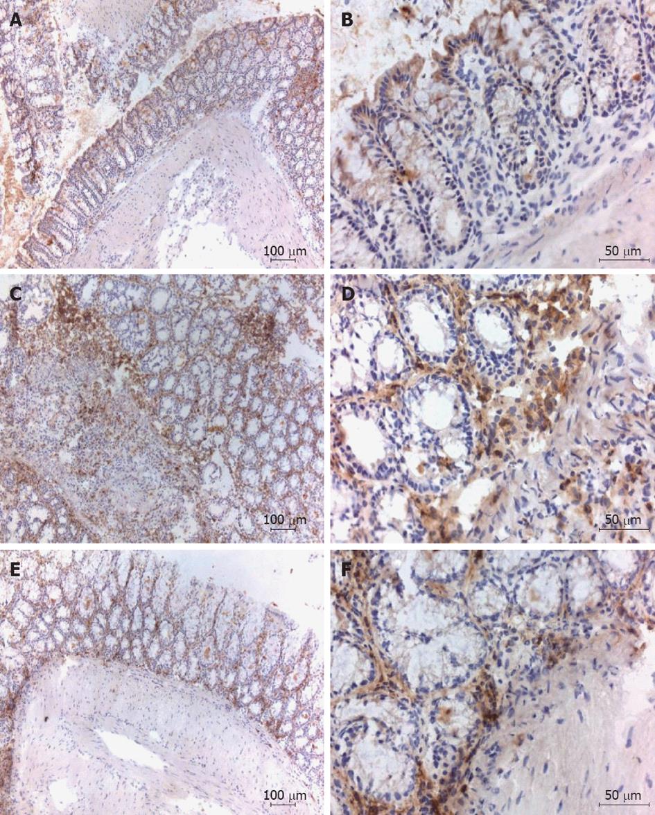
Figure 5 Immunohistochemistry pictures depicting CD4 staining colon tissue.
A, B: Healthy control mice; C, D: Diseased control mice; E, F: Adeno associated virus (AAV) 5 [cytomegalovirus (CMV) promoter T-cell epitopes (Tregitope) 167] pre-treated mice. Specific immunohistochemical staining showed inflammatory cell infiltrates present in trinitrobenzene sulfonate (TNBS) treated mice with or without administration with Tregitope 167 consisted of CD4 positive cells, localized in the sub-epithelial layer, in the lamina propria (C-F) and for the diseased control also in the muscular layer (C,D). Depicted are representative data from a single mouse.
- Citation: van der Marel S, Majowicz A, Kwikkers K, van Logtenstein R, te Velde AA, De Groot AS, Meijer SL, van Deventer SJ, Petry H, Hommes DW, Ferreira V. Adeno-associated virus mediated delivery of Tregitope 167 ameliorates experimental colitis. World J Gastroenterol 2012; 18(32): 4288-4299
- URL: https://www.wjgnet.com/1007-9327/full/v18/i32/4288.htm
- DOI: https://dx.doi.org/10.3748/wjg.v18.i32.4288


