Copyright
©2012 Baishideng Publishing Group Co.
World J Gastroenterol. Aug 7, 2012; 18(29): 3917-3920
Published online Aug 7, 2012. doi: 10.3748/wjg.v18.i29.3917
Published online Aug 7, 2012. doi: 10.3748/wjg.v18.i29.3917
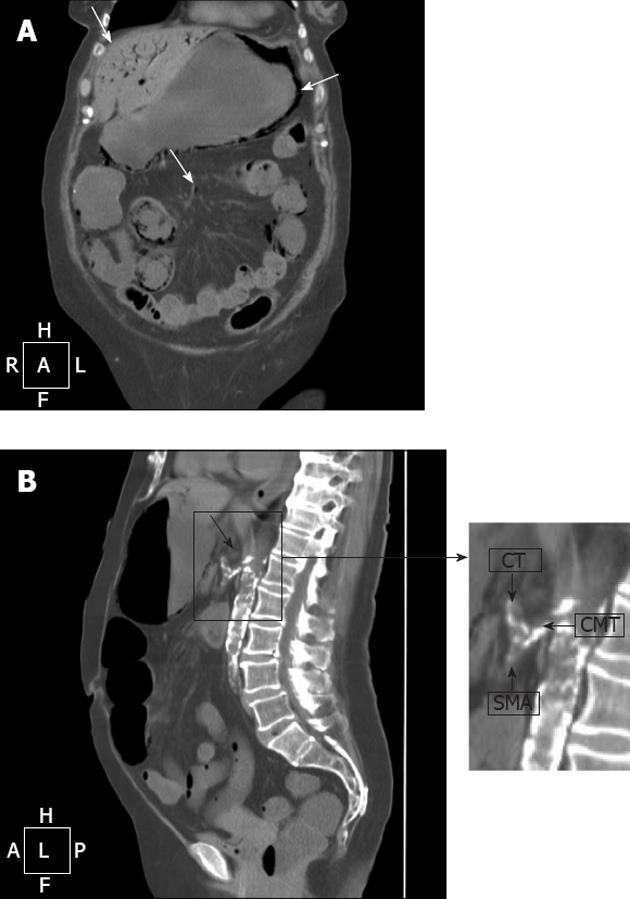
Figure 2 Computed tomography of the abdomen.
A: Pneumatosis of the wall of stomach and small bowel (arrow in the right). Intraparenchimal air in ventral portions of the left liver and fourth segment (arrow in the left); air within the branches of the mesenteric vein (arrow in the centre); B:Thrombosis of the celiacomesenteric trunk (CMT) (arrow). In the detail: common origin of celiac trunk (CT) and superior mesenteric artery (SMA) from the CMT.
- Citation: Lovisetto F, Finocchiaro De Lorenzi G, Stancampiano P, Corradini C, De Cesare F, Geraci O, Manzi M, Arceci F. Thrombosis of celiacomesenteric trunk: Report of a case. World J Gastroenterol 2012; 18(29): 3917-3920
- URL: https://www.wjgnet.com/1007-9327/full/v18/i29/3917.htm
- DOI: https://dx.doi.org/10.3748/wjg.v18.i29.3917


