Copyright
©2012 Baishideng Publishing Group Co.
World J Gastroenterol. May 28, 2012; 18(20): 2586-2590
Published online May 28, 2012. doi: 10.3748/wjg.v18.i20.2586
Published online May 28, 2012. doi: 10.3748/wjg.v18.i20.2586
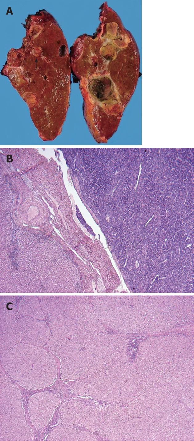
Figure 3 Macroscopic and microscopic appearance of the resected liver.
A: A tumor thrombus was detected in the right portal vein (arrow); B: A moderately differentiated hepatocellular carcinoma with a trabecular pattern was seen in the right portal vein [hematoxylin and eosin (HE) stain, ×40]; C: Fibrosis of varying extent was observed in the cancer-free area. Some pseudolobules with severe fibrosis were present in the left part of the photograph (HE stain, ×40).
- Citation: Honda K, Seike M, Maehara SI, Tahara K, Anai H, Moriuchi A, Muro T. Lamivudine treatment enabling right hepatectomy for hepatocellular carcinoma in decompensated cirrhosis. World J Gastroenterol 2012; 18(20): 2586-2590
- URL: https://www.wjgnet.com/1007-9327/full/v18/i20/2586.htm
- DOI: https://dx.doi.org/10.3748/wjg.v18.i20.2586


