Copyright
©2012 Baishideng Publishing Group Co.
World J Gastroenterol. May 28, 2012; 18(20): 2586-2590
Published online May 28, 2012. doi: 10.3748/wjg.v18.i20.2586
Published online May 28, 2012. doi: 10.3748/wjg.v18.i20.2586
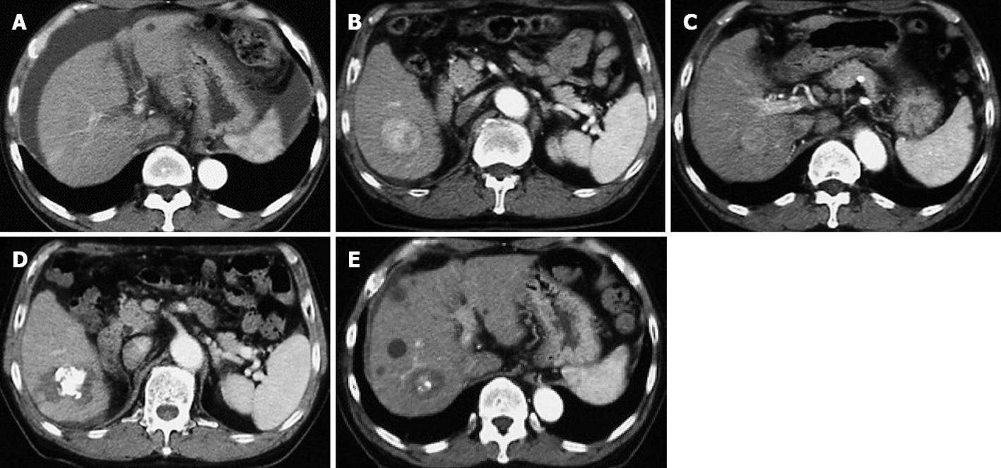
Figure 1 Computed tomography in our patient.
It shows a cirrhotic pattern of the liver and massive ascites at first admission (A); Dynamic computed tomography revealed two hepatocellular carcinomas, 4.5 cm (B) and 2.5 cm (C) in diameter, in the right lobe; These two lesions were treated by transcatheter arterial chemoembolization and radiofrequency ablation (D, E).
- Citation: Honda K, Seike M, Maehara SI, Tahara K, Anai H, Moriuchi A, Muro T. Lamivudine treatment enabling right hepatectomy for hepatocellular carcinoma in decompensated cirrhosis. World J Gastroenterol 2012; 18(20): 2586-2590
- URL: https://www.wjgnet.com/1007-9327/full/v18/i20/2586.htm
- DOI: https://dx.doi.org/10.3748/wjg.v18.i20.2586


