Copyright
©2012 Baishideng Publishing Group Co.
World J Gastroenterol. May 14, 2012; 18(18): 2291-2294
Published online May 14, 2012. doi: 10.3748/wjg.v18.i18.2291
Published online May 14, 2012. doi: 10.3748/wjg.v18.i18.2291
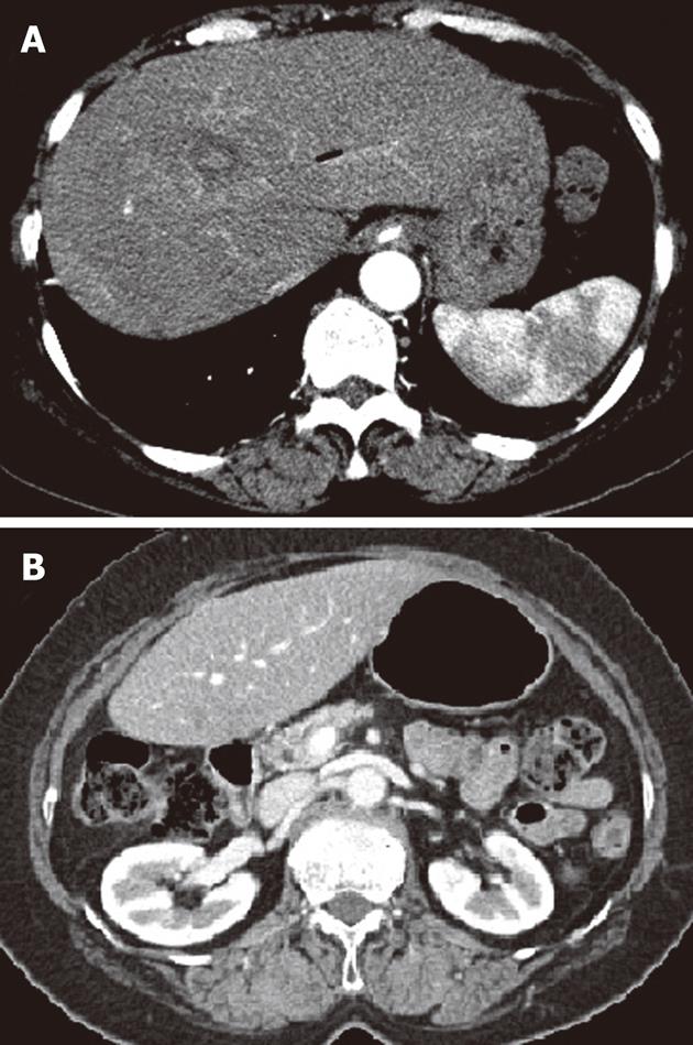
Figure 5 Follow-up of abdominal computed tomography scans 3 mo later showed that both previously noted hepatic artery pseudoaneurysm in the left lateral segment (A) and diffuse swelling and infiltration of pancreas (B) had disappeared.
- Citation: Yu YH, Sohn JH, Kim TY, Jeong JY, Han DS, Jeon YC, Kim MY. Hepatic artery pseudoaneurysm caused by acute idiopathic pancreatitis. World J Gastroenterol 2012; 18(18): 2291-2294
- URL: https://www.wjgnet.com/1007-9327/full/v18/i18/2291.htm
- DOI: https://dx.doi.org/10.3748/wjg.v18.i18.2291


