Copyright
©2012 Baishideng Publishing Group Co.
World J Gastroenterol. May 14, 2012; 18(18): 2291-2294
Published online May 14, 2012. doi: 10.3748/wjg.v18.i18.2291
Published online May 14, 2012. doi: 10.3748/wjg.v18.i18.2291
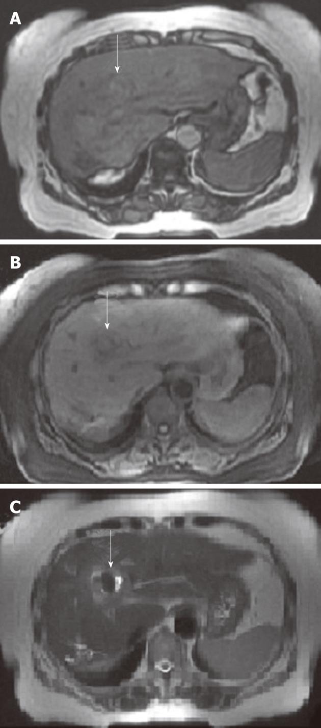
Figure 4 Magnetic resonance imaging scans of the abdomen revealed that hepatic artery pseudoaneurysm was replaced by thrombus formation (white arrows) in the left lateral segment in-phase (A) and out-of-phase (B) T1-weighted images and T2-weighted image (C).
- Citation: Yu YH, Sohn JH, Kim TY, Jeong JY, Han DS, Jeon YC, Kim MY. Hepatic artery pseudoaneurysm caused by acute idiopathic pancreatitis. World J Gastroenterol 2012; 18(18): 2291-2294
- URL: https://www.wjgnet.com/1007-9327/full/v18/i18/2291.htm
- DOI: https://dx.doi.org/10.3748/wjg.v18.i18.2291


