Copyright
©2012 Baishideng Publishing Group Co.
World J Gastroenterol. May 14, 2012; 18(18): 2291-2294
Published online May 14, 2012. doi: 10.3748/wjg.v18.i18.2291
Published online May 14, 2012. doi: 10.3748/wjg.v18.i18.2291
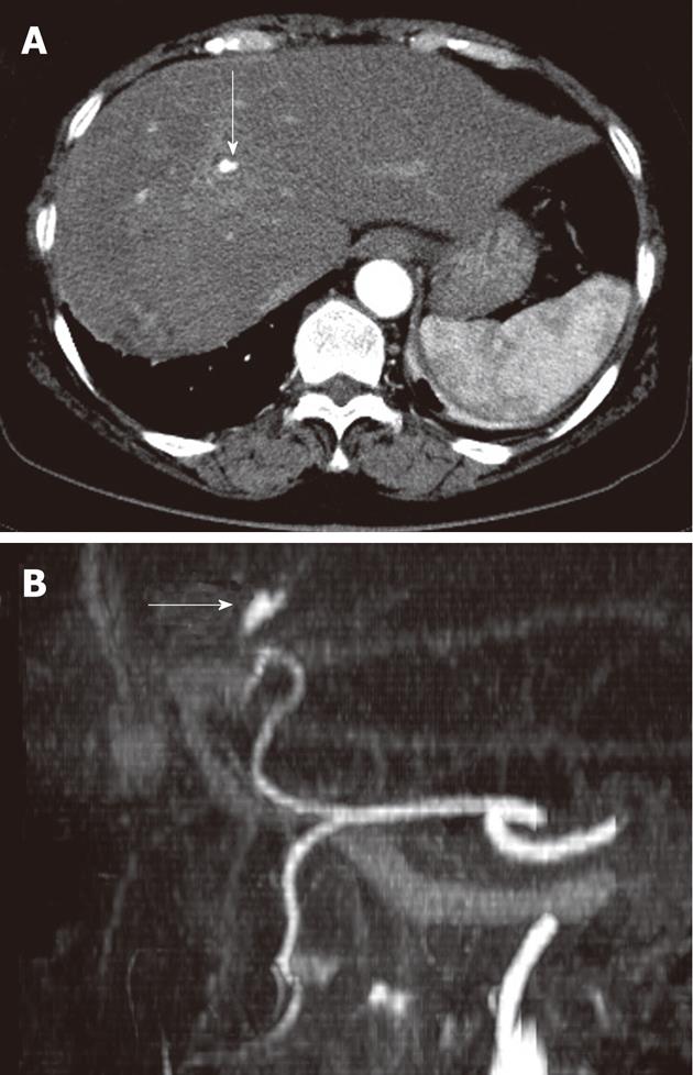
Figure 2 Follow up of abdominal computed tomography scan (A) due to abruptly aggravated abdominal pain and reconstructed computed tomography angiogram (B) showed newly developed hepatic artery pseudoaneurysm in the left lateral segment of liver (white arrows).
- Citation: Yu YH, Sohn JH, Kim TY, Jeong JY, Han DS, Jeon YC, Kim MY. Hepatic artery pseudoaneurysm caused by acute idiopathic pancreatitis. World J Gastroenterol 2012; 18(18): 2291-2294
- URL: https://www.wjgnet.com/1007-9327/full/v18/i18/2291.htm
- DOI: https://dx.doi.org/10.3748/wjg.v18.i18.2291


