Copyright
©2012 Baishideng Publishing Group Co.
World J Gastroenterol. Apr 7, 2012; 18(13): 1538-1544
Published online Apr 7, 2012. doi: 10.3748/wjg.v18.i13.1538
Published online Apr 7, 2012. doi: 10.3748/wjg.v18.i13.1538
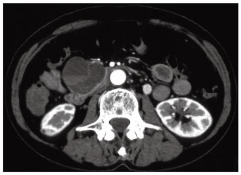
Figure 1 Contrast-enhanced computed tomography scan obtained in the arterial phase showing a multilocular cystic mass in the uncinate process of the pancreas.
No pancreatic ductal dilatation or invasion into adjacent arteries or portal vein are identified.
- Citation: Moriya T, Kimura W, Hirai I, Takeshita A, Tezuka K, Watanabe T, Mizutani M, Fuse A. Pancreatic schwannoma: Case report and an updated 30-year review of the literature yielding 47 cases. World J Gastroenterol 2012; 18(13): 1538-1544
- URL: https://www.wjgnet.com/1007-9327/full/v18/i13/1538.htm
- DOI: https://dx.doi.org/10.3748/wjg.v18.i13.1538


