Copyright
©2011 Baishideng Publishing Group Co.
World J Gastroenterol. Dec 14, 2011; 17(46): 5110-5116
Published online Dec 14, 2011. doi: 10.3748/wjg.v17.i46.5110
Published online Dec 14, 2011. doi: 10.3748/wjg.v17.i46.5110
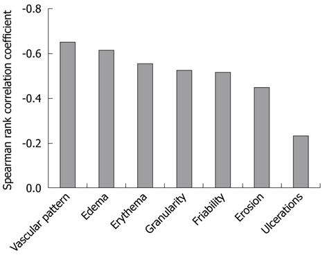
Figure 4 Comparison of seven endoscopic ulcerative colitis features on white light imaging and the green color component on autofluorescence imaging.
The green color component on autofluorescence imaging is well correlated with vascular pattern (r = -0.65, P < 0.01) and edema (r = -0.62, P < 0.01) scores on WLI, but not with the ulcer score (r = -0.23, P < 0.01) (Spearman rank correlation coefficient). WLI: White light imaging.
- Citation: Osada T, Arakawa A, Sakamoto N, Ueyama H, Shibuya T, Ogihara T, Yao T, Watanabe S. Autofluorescence imaging endoscopy for identification and assessment of inflammatory ulcerative colitis. World J Gastroenterol 2011; 17(46): 5110-5116
- URL: https://www.wjgnet.com/1007-9327/full/v17/i46/5110.htm
- DOI: https://dx.doi.org/10.3748/wjg.v17.i46.5110


