Copyright
©2011 Baishideng Publishing Group Co.
World J Gastroenterol. Dec 14, 2011; 17(46): 5110-5116
Published online Dec 14, 2011. doi: 10.3748/wjg.v17.i46.5110
Published online Dec 14, 2011. doi: 10.3748/wjg.v17.i46.5110
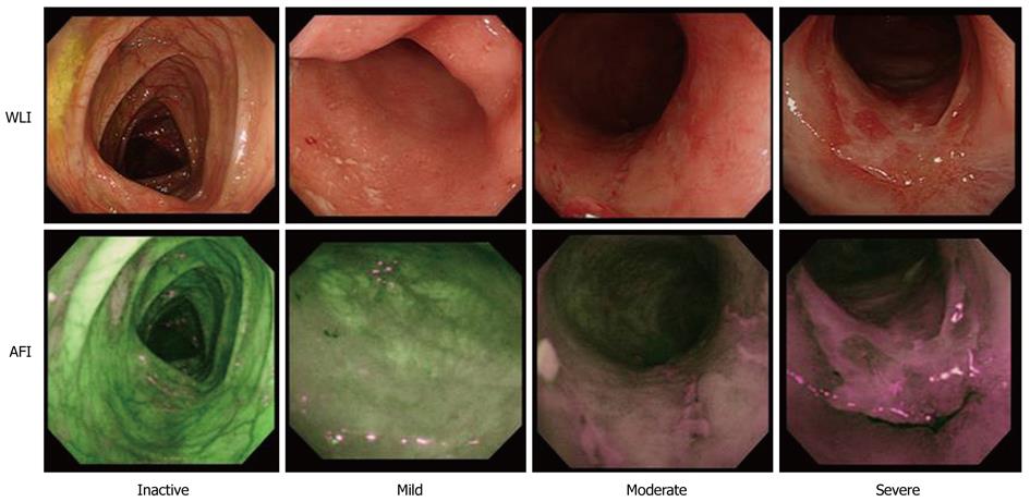
Figure 2 Representative endoscopic photographs of ulcerative colitis using white light imaging (upper row) and autofluorescence imaging (lower row) at the same sites according to the level of endoscopic ulcerative colitis activity (inactive, mild, moderate and severe).
The color of the large intestinal mucosa on the autofluorescence imaging (AFI) changes by degrees, from green to grayish and magenta color. WLI: White light imaging.
- Citation: Osada T, Arakawa A, Sakamoto N, Ueyama H, Shibuya T, Ogihara T, Yao T, Watanabe S. Autofluorescence imaging endoscopy for identification and assessment of inflammatory ulcerative colitis. World J Gastroenterol 2011; 17(46): 5110-5116
- URL: https://www.wjgnet.com/1007-9327/full/v17/i46/5110.htm
- DOI: https://dx.doi.org/10.3748/wjg.v17.i46.5110


