Copyright
©2011 Baishideng Publishing Group Co.
World J Gastroenterol. Dec 7, 2011; 17(45): 5028-5031
Published online Dec 7, 2011. doi: 10.3748/wjg.v17.i45.5028
Published online Dec 7, 2011. doi: 10.3748/wjg.v17.i45.5028
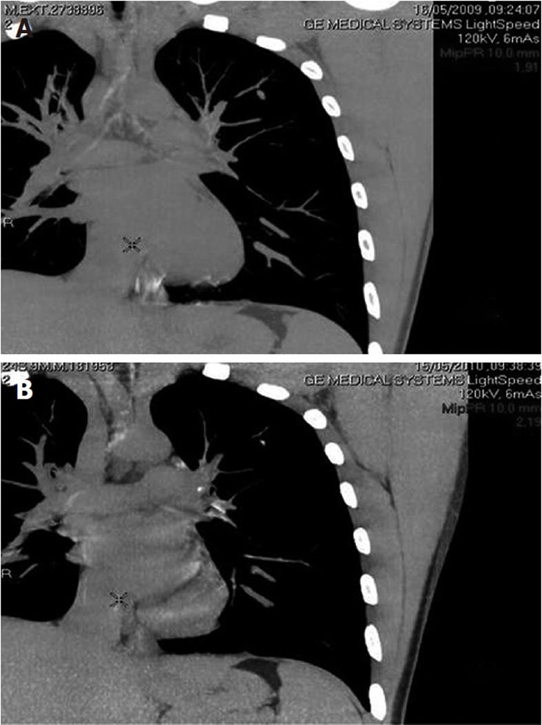
Figure 2 Chest computed tomography.
A: Small nodule, measuring 0.8 to 0.6 cm, in the upper lobe of the left lung; B: Small calcified nodule located in the apico-posterior lobe of the left lung, measuring 0.5 cm in diameter.
- Citation: Leite MR, Santos SS, Lyra AC, Mota J, Santana GO. Thalidomide induces mucosal healing in Crohn's disease: Case report. World J Gastroenterol 2011; 17(45): 5028-5031
- URL: https://www.wjgnet.com/1007-9327/full/v17/i45/5028.htm
- DOI: https://dx.doi.org/10.3748/wjg.v17.i45.5028


