Copyright
©2011 Baishideng Publishing Group Co.
World J Gastroenterol. Jun 21, 2011; 17(23): 2801-2811
Published online Jun 21, 2011. doi: 10.3748/wjg.v17.i23.2801
Published online Jun 21, 2011. doi: 10.3748/wjg.v17.i23.2801
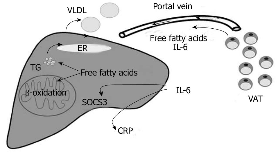
Figure 1 Crosstalk of visceral adipose tissue and the liver.
Free fatty acids and Interleukin (IL)-6 released by visceral adipose tissue (VAT) are transported to the liver by the portal vein. Free fatty acids promote steatosis, enhance β-oxidation and the release of very low density lipoproteins (VLDL) contributing to dyslipidemia. IL-6 induces hepatic C-reactive protein (CRP) synthesis and suppressor of cytokine signalling 3 (SOCS3), and thereby is linked to systemic inflammation and hepatic insulin resistance, respectively (adapted from Schaffler A, Scholmerich J, Buchler C. Mechanisms of disease: adipocytokines and visceral adipose tissue--emerging role in nonalcoholic fatty liver disease. Nat Clin Pract Gastroenterol Hepatol 2005; 2: 273-280[6]). ER: Endoplasmatic reticulum; TG: Triglycerides.
- Citation: Buechler C, Wanninger J, Neumeier M. Adiponectin, a key adipokine in obesity related liver diseases. World J Gastroenterol 2011; 17(23): 2801-2811
- URL: https://www.wjgnet.com/1007-9327/full/v17/i23/2801.htm
- DOI: https://dx.doi.org/10.3748/wjg.v17.i23.2801


