Copyright
©2011 Baishideng Publishing Group Co.
World J Gastroenterol. Jan 14, 2011; 17(2): 226-230
Published online Jan 14, 2011. doi: 10.3748/wjg.v17.i2.226
Published online Jan 14, 2011. doi: 10.3748/wjg.v17.i2.226
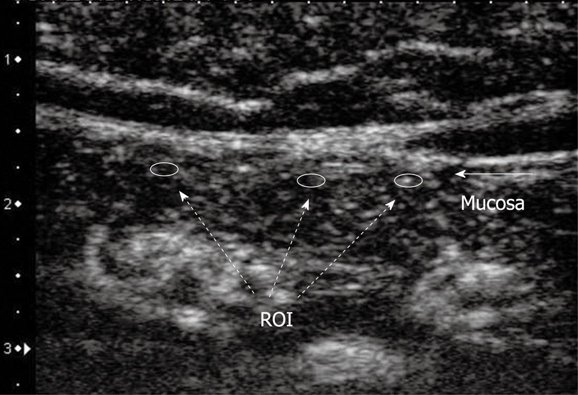
Figure 2 Image of contrast-enhanced ultrasonography.
Regions of interests (ROI) were placed in the mucosal area of the small bowel at three regions. A time intensity curve of blood flow enhancement signal was plotted from recorded ultrasonographic images.
- Citation: Nishida U, Kato M, Nishida M, Kamada G, Yoshida T, Ono S, Shimizu Y, Asaka M. Evaluation of small bowel blood flow in healthy subjects receiving low-dose aspirin. World J Gastroenterol 2011; 17(2): 226-230
- URL: https://www.wjgnet.com/1007-9327/full/v17/i2/226.htm
- DOI: https://dx.doi.org/10.3748/wjg.v17.i2.226


