Copyright
©2011 Baishideng Publishing Group Co.
World J Gastroenterol. Mar 28, 2011; 17(12): 1584-1593
Published online Mar 28, 2011. doi: 10.3748/wjg.v17.i12.1584
Published online Mar 28, 2011. doi: 10.3748/wjg.v17.i12.1584
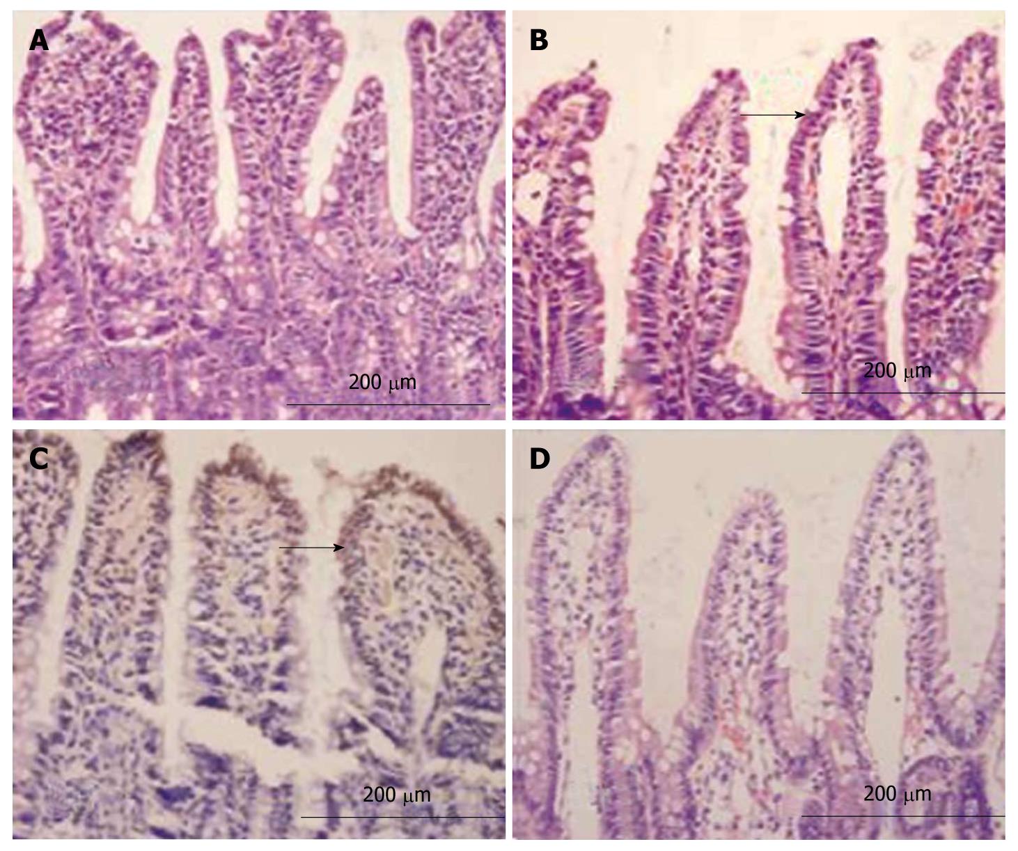
Figure 2 Light microscopy.
Smooth intestinal mucosa with intact epithelia and ordered villi in group C (A), exfoliated and incomplete intestinal mucosa with thickened mucosa and reduced villi accompanying irregular morphology and disorganized villous epithelia in group H (B), atrophic and thinned villi accompanying a loose and disordered arrangement as well as edema and infiltration of inflammatory cells in mastoid lamina of villi and lodged and exfoliated villi with loss of goblet cells and red blood cell effusion around the capillaries in group HH (C), relatively intact intestinal mucosal villi with ordered arrangement and alleviated edema in mastoid lamina of villi accompanying a few infiltrated inflammatory cells in group HG (D) (HE, × 200).
- Citation: Zhou QQ, Yang DZ, Luo YJ, Li SZ, Liu FY, Wang GS. Over-starvation aggravates intestinal injury and promotes bacterial and endotoxin translocation under high-altitude hypoxic environment. World J Gastroenterol 2011; 17(12): 1584-1593
- URL: https://www.wjgnet.com/1007-9327/full/v17/i12/1584.htm
- DOI: https://dx.doi.org/10.3748/wjg.v17.i12.1584


