Copyright
©2011 Baishideng Publishing Group Co.
World J Gastroenterol. Mar 21, 2011; 17(11): 1442-1447
Published online Mar 21, 2011. doi: 10.3748/wjg.v17.i11.1442
Published online Mar 21, 2011. doi: 10.3748/wjg.v17.i11.1442
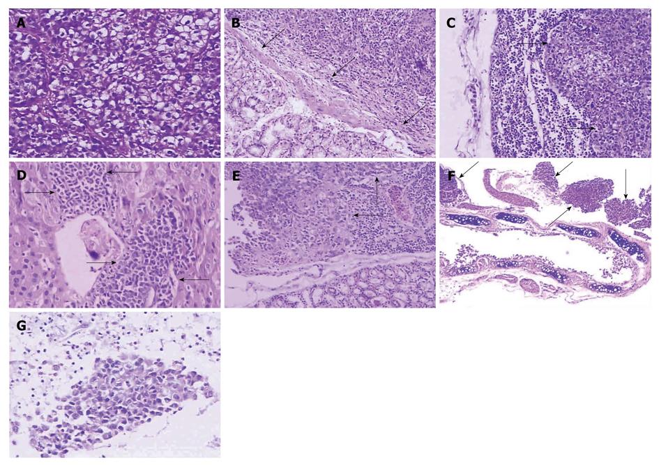
Figure 2 Histopathologic structure of SGC-7901 tumors by HE staining.
A: Tumor cells showed nuclear polymorphism, nuclear hyperchromatism, and much mucus in the cytoplasm (× 200); B: The primary tumor destroyed muscularis mucosae (arrows) (× 100); C: The lymph nodes were infiltrated by metastatic tumor (arrows) (× 100); D: Metastases (arrows) infiltrated into the liver (× 200); E: Metastatic tumor (arrows) invaded the kidney (× 100); F: Tumor (arrows) metastasized to the lung and surrounded bronchia or bronchiole (× 40); G: Cast-off tumor cells were detected in ascites (× 200).
- Citation: Li Y, Li B, Zhang Y, Xiang CP, Li YY, Wu XL. Serial observations on an orthotopic gastric cancer model constructed using improved implantation technique. World J Gastroenterol 2011; 17(11): 1442-1447
- URL: https://www.wjgnet.com/1007-9327/full/v17/i11/1442.htm
- DOI: https://dx.doi.org/10.3748/wjg.v17.i11.1442


