Copyright
©2011 Baishideng Publishing Group Co.
World J Gastroenterol. Mar 21, 2011; 17(11): 1383-1399
Published online Mar 21, 2011. doi: 10.3748/wjg.v17.i11.1383
Published online Mar 21, 2011. doi: 10.3748/wjg.v17.i11.1383
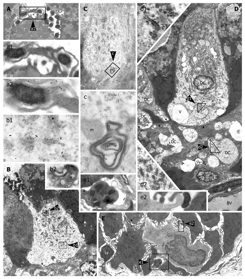
Figure 3 Intraepithelial dendritic cells in Helicobacter pylori-positive human gastric biopsies with active inflammation.
A (5000 ×): Luminal ending of a dendritic cell (DC) process abutting on a collection of Helicobacter pylori (H. pylori); enlarged in a1 (16 000 ×) to show envelopment of a bacterium by caliceal veils (right); note in a2 (28 000 ×; detail of a1), close adherence of a clubbed process (bottom left) to another bacterium showing VacA immunoreactivity of outer membrane and flagella (upper right); B (3000 ×): Intraepithelial DC with a narrow luminal process directly contacting bacteria; note close membrane adhesion to surrounding epithelial cells, focal interruption of the basal lamina (arrow), and outer membrane protein (OMP) immunoreactivity of cytoplasmic vacuoles (b1, 32 000 ×) and a multivesicular late endosome/lysosome body (b2, 22 000 ×); C (8000 ×): DC with clear cytoplasm, close adherence to surrounding epithelial cells, numerous mitochondria, sparse ribosomes and small rough endoplasmic reticulum (RER) cisternae; cytoplasmic vesicles and membranous remnants of an intracellular bacterium, enlarged in c (42 000 ×), show VacA immunoreactivity; D (2000 ×): Nucleated DC with abundant supranuclear mitochondria, no secretory granules, small tubular RER cisternae (enlarged in d1, 18 000 ×), several cytoplasmic vesicles, scattered vacuoles (v), and close adhesion to epithelial cells. On the left, two DC processes (arrows) are contacting the basal membrane (arrowheads). Several cross or longitudinal sections of partly swollen cell processes are observed in the lamina propria; the largest of which (enlarged in d2, 12 000 ×) shows ultrastructural homology with DC cytoplasm, including scattered, small RER cisternae and vesicles; the precise cells of origin of such lamina propria processes could not be assessed. BV: Blood vessel; fg: Foveolar granules; E (2000 ×): Base of foveolar epithelium showing an immature monocytoid cell (DC precursor?) with kidney-shaped nucleus, scattered ribosomes, a few juxtanuclear lysosomes, and no vesicles or granules; note a caliceal process embracing a mast cell (enlarged in e1, 15 000 ×; scroll bodies, arrow), a clubbed process adhering to H. pylori (enlarged in e2, 55 000 ×), a lymphoid cell (Ly) crossing the epithelium basal membrane and another intercellular bacterium (arrow). Reprinted from Necchi et al[73], with permission from John Wiley.
- Citation: Ricci V, Romano M, Boquet P. Molecular cross-talk between Helicobacter pylori and human gastric mucosa. World J Gastroenterol 2011; 17(11): 1383-1399
- URL: https://www.wjgnet.com/1007-9327/full/v17/i11/1383.htm
- DOI: https://dx.doi.org/10.3748/wjg.v17.i11.1383


