Copyright
©2011 Baishideng Publishing Group Co.
World J Gastroenterol. Mar 21, 2011; 17(11): 1383-1399
Published online Mar 21, 2011. doi: 10.3748/wjg.v17.i11.1383
Published online Mar 21, 2011. doi: 10.3748/wjg.v17.i11.1383
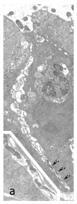
Figure 1 Helicobacter pylori penetration in human gastric epithelium in vivo.
Three Helicobacter pylori (H. pylori) organisms (enlarged in a; 16 800 ×) lying in the deep intercellular intraepithelial space, just above the basal membrane. Note also luminal bacteria (top) overlying an apparently preserved tight junction, dilation of the underlying intercellular space, filled with lateral membrane plications, and an intraepithelial granulocyte (middle right, 6300 ×). Reprinted from Necchi et al[57], with permission from Elsevier.
- Citation: Ricci V, Romano M, Boquet P. Molecular cross-talk between Helicobacter pylori and human gastric mucosa. World J Gastroenterol 2011; 17(11): 1383-1399
- URL: https://www.wjgnet.com/1007-9327/full/v17/i11/1383.htm
- DOI: https://dx.doi.org/10.3748/wjg.v17.i11.1383


