Copyright
©2011 Baishideng Publishing Group Co.
World J Gastroenterol. Jan 7, 2011; 17(1): 130-134
Published online Jan 7, 2011. doi: 10.3748/wjg.v17.i1.130
Published online Jan 7, 2011. doi: 10.3748/wjg.v17.i1.130
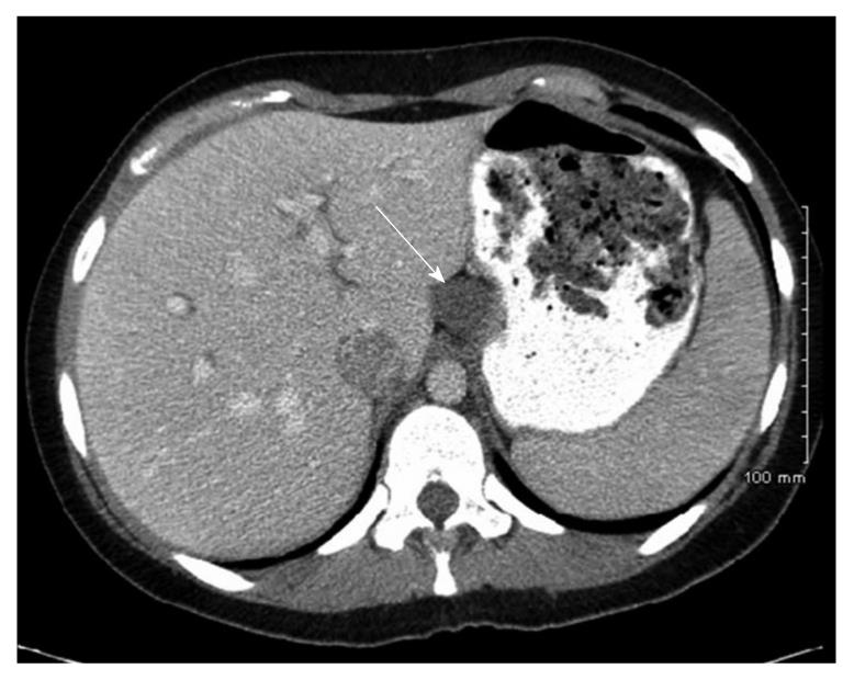
Figure 4 Case 2.
Computed tomography-scan of the abdomen and pelvis with intravenous and oral contrast showing a non-enhancing cystic structure (arrow) abutting the lesser curvature of the stomach and liver along the ligamentum venosum.
- Citation: Khoury T, Rivera L. Foregut duplication cysts: A report of two cases with emphasis on embryogenesis. World J Gastroenterol 2011; 17(1): 130-134
- URL: https://www.wjgnet.com/1007-9327/full/v17/i1/130.htm
- DOI: https://dx.doi.org/10.3748/wjg.v17.i1.130


