Copyright
©2011 Baishideng Publishing Group Co.
World J Gastroenterol. Jan 7, 2011; 17(1): 130-134
Published online Jan 7, 2011. doi: 10.3748/wjg.v17.i1.130
Published online Jan 7, 2011. doi: 10.3748/wjg.v17.i1.130
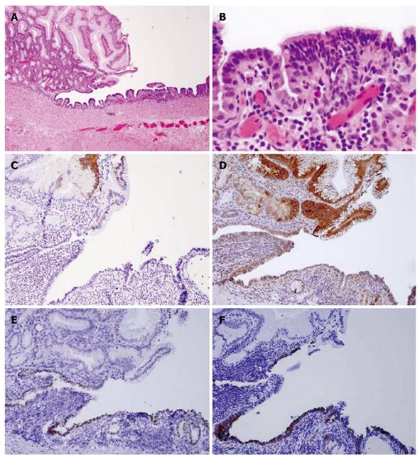
Figure 3 Case 1.
A: Hematoxylin and eosin satin of gastric/ciliated epithelium (4 ×); B: High power view of the ciliated epithelium (40 ×); C: CK20 showing staining in the surface gastric epithelium, but not in the ciliated epithelium (10 ×); D: MUC5a/c staining showing positive expression in the gastric epithelium but not in the ciliated epithelium (10 ×); E: Thyroid transcription factor-1 nuclear staining in the ciliated epithelium only (10 ×); F: Surfactant staining in the ciliated epithelium only (10 ×).
- Citation: Khoury T, Rivera L. Foregut duplication cysts: A report of two cases with emphasis on embryogenesis. World J Gastroenterol 2011; 17(1): 130-134
- URL: https://www.wjgnet.com/1007-9327/full/v17/i1/130.htm
- DOI: https://dx.doi.org/10.3748/wjg.v17.i1.130


