Copyright
©2010 Baishideng Publishing Group Co.
World J Gastroenterol. Nov 14, 2010; 16(42): 5306-5316
Published online Nov 14, 2010. doi: 10.3748/wjg.v16.i42.5306
Published online Nov 14, 2010. doi: 10.3748/wjg.v16.i42.5306
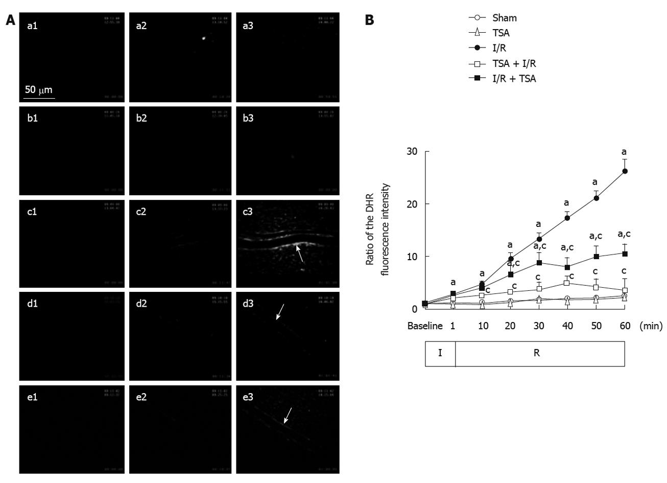
Figure 3 The effect of pre-treatment and post-treatment of total salvianolic acid on ischemia-reperfusion-induced dihydrorhodamine 123 fluorescence in rat mesenteric venular wall.
A: Representative images of the changes in fluorescence intensity of the H2O2-sensitive probe dihydrorhodamine 123 (DHR) in the rat mesenteric venular wall. a1-a3: DHR fluorescence images of rat mesentery of Sham group at baseline, 10 and 60 min, respectively. b1-b3: Fluorescence images of rat mesentery of total salvianolic acid (TSA) group at baseline, 10 and 60 min, respectively. c1-c3: Fluorescence images of rat mesentery of ischemia-reperfusion (I/R) group at baseline, 10 and 60 min, respectively. d1-d3: Fluorescence images of rat mesentery of TSA + I/R group at baseline, 10 and 60 min, respectively. e1-e3: Fluorescence images of rat mesentery of I/R + TSA group at baseline, 10 and 60 min, respectively. Baseline, no DHR fluorescence is visible in all groups (a1-e1). 60 min after I/R, prominent DHR fluorescence occurs on the wall of venule of rat mesentery (c3-e3). Arrows: DHR fluorescence on the venular wall; B: Time course of changes in DHR fluorescence ratio on the venular walls. Sham: Sham group; TSA: TSA group; I/R: I/R group; TSA + I/R: TSA plus I/R group; I/R + TSA: I/R plus TSA group. Data was expressed as mean ± SE of six animals. aP < 0.05 vs sham group; cP < 0.05 vs I/R alone.
- Citation: Wang MX, Liu YY, Hu BH, Wei XH, Chang X, Sun K, Fan JY, Liao FL, Wang CS, Zheng J, Han JY. Total salvianolic acid improves ischemia-reperfusion-induced microcirculatory disturbance in rat mesentery. World J Gastroenterol 2010; 16(42): 5306-5316
- URL: https://www.wjgnet.com/1007-9327/full/v16/i42/5306.htm
- DOI: https://dx.doi.org/10.3748/wjg.v16.i42.5306


