Copyright
©2010 Baishideng.
World J Gastroenterol. Jan 21, 2010; 16(3): 281-288
Published online Jan 21, 2010. doi: 10.3748/wjg.v16.i3.281
Published online Jan 21, 2010. doi: 10.3748/wjg.v16.i3.281
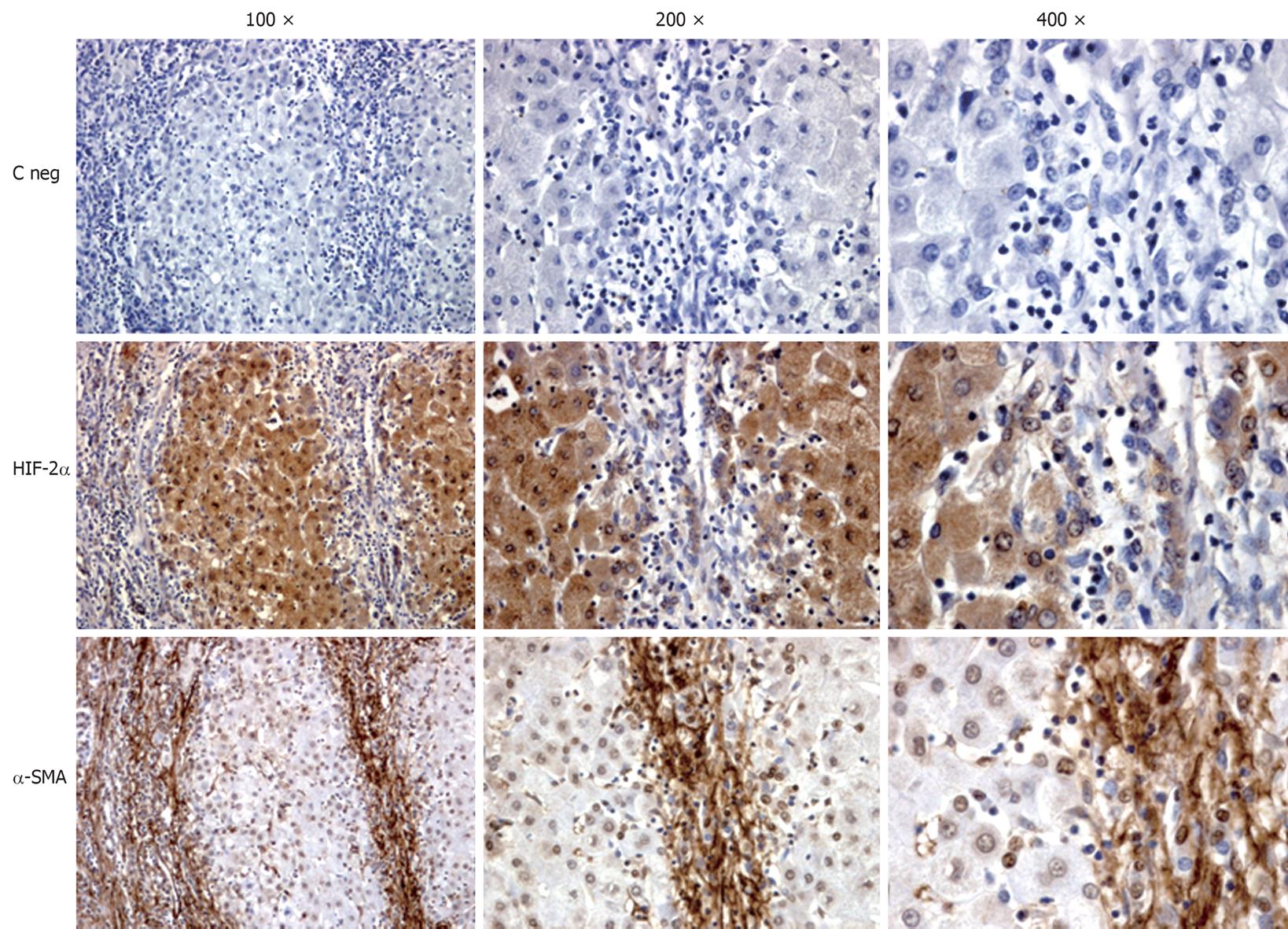
Figure 1 Immunohistochemical analysis performed on paraffin liver sections from patients with hepatitis C virus (HCV)-related liver cirrhosis (METAVIR F4).
Sections (2 μm thick) were incubated with specific antibodies raised against HIF-2α or α-SMA that positively stain cells exposed to hypoxia (HIF-2α) or myofibroblast-like cells (α-SMA). Primary antibodies were labelled by using EnVision, HRP-labelled System (DAKO) antibodies and visualized by 3’-diaminobenzidine substrate. Negative controls (C neg) were obtained by replacing the respective primary antibodies by isotype and concentrations matched irrelevant antibody. Original magnification is indicated.
- Citation: Paternostro C, David E, Novo E, Parola M. Hypoxia, angiogenesis and liver fibrogenesis in the progression of chronic liver diseases. World J Gastroenterol 2010; 16(3): 281-288
- URL: https://www.wjgnet.com/1007-9327/full/v16/i3/281.htm
- DOI: https://dx.doi.org/10.3748/wjg.v16.i3.281


