Copyright
©2010 Baishideng.
World J Gastroenterol. May 28, 2010; 16(20): 2571-2576
Published online May 28, 2010. doi: 10.3748/wjg.v16.i20.2571
Published online May 28, 2010. doi: 10.3748/wjg.v16.i20.2571
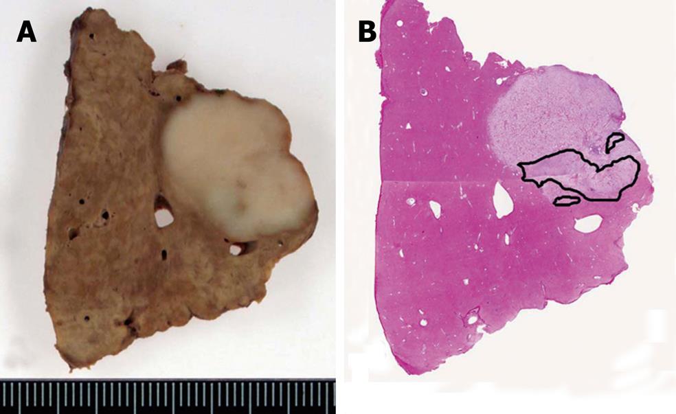
Figure 3 Tumor images.
A: Photograph of the cut surface of the tumor fixed in formalin shows a yellowish-white, solid tumor, measuring 1.5 cm × 2.2 cm. Although the tumor margin is clear, there is no fibrous capsule. Neither bleeding nor blood vessels inside the tumor are observed. The outline margin with the surrounding liver is irregular; B: Scanning image. The tumor consists of clear cells. The margin is clear. The area surrounded by the black line is the poorly differentiated area.
- Citation: Toriyama E, Nanashima A, Hayashi H, Abe K, Kinoshita N, Yuge S, Nagayasu T, Uetani M, Hayashi T. A case of intrahepatic clear cell cholangiocarcinoma. World J Gastroenterol 2010; 16(20): 2571-2576
- URL: https://www.wjgnet.com/1007-9327/full/v16/i20/2571.htm
- DOI: https://dx.doi.org/10.3748/wjg.v16.i20.2571


